. 2015 Mar 3;10(3):e0118415.
doi: 10.1371/journal.pone.0118415. eCollection 2015.

Function and evolutionary origin of unicellular camera-type eye structure

Affiliations

Function and evolutionary origin of unicellular camera-type eye structure

Shiho Hayakawa et al. PLoS One. .

Abstract

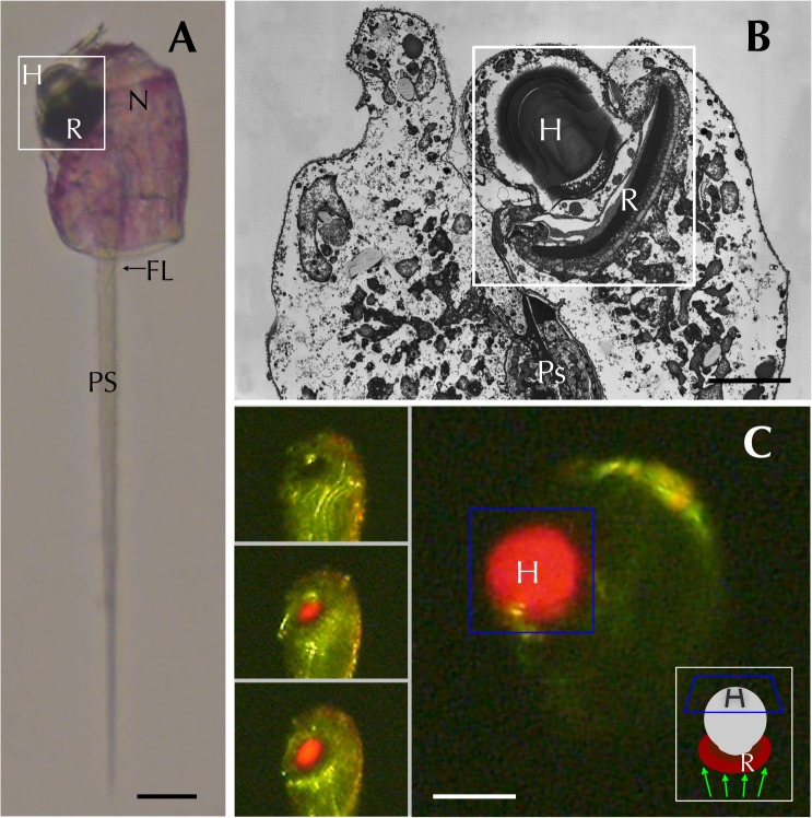
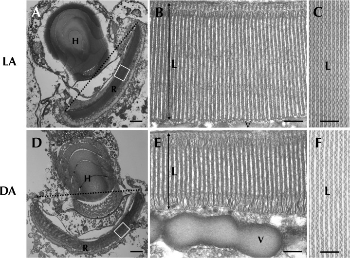
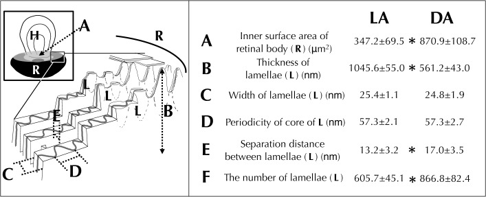
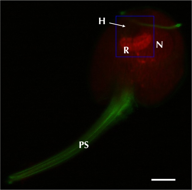
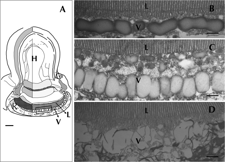
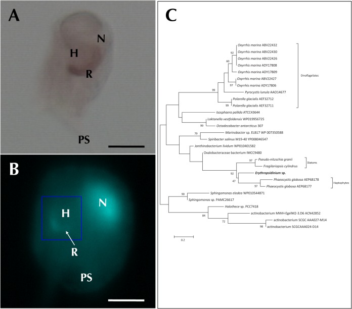
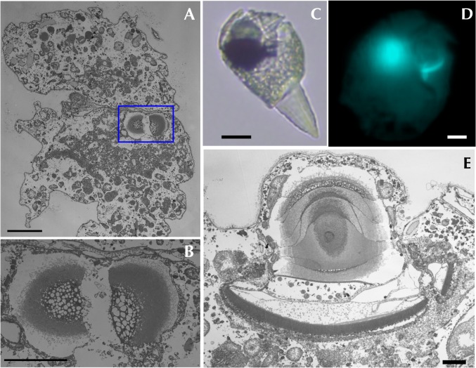
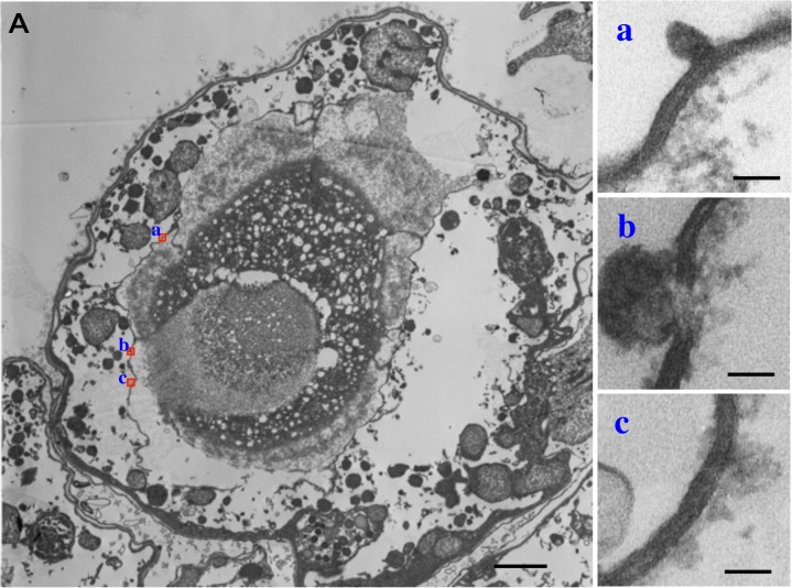
The ocelloid is an extraordinary eyespot organelle found only in the dinoflagellate family Warnowiaceae. It contains retina- and lens-like structures called the retinal body and the hyalosome. The ocelloid has been an evolutionary enigma because of its remarkable resemblance to the multicellular camera-type eye. To determine if the ocelloid is functionally photoreceptive, we investigated the warnowiid dinoflagellate Erythropsidinium. Here, we show that the morphology of the retinal body changed depending on different illumination conditions and the hyalosome manifests the refractile nature. Identifying a rhodopsin gene fragment in Erythropsidinium ESTs that is expressed in the retinal body by in situ hybridization, we also show that ocelloids are actually light sensitive photoreceptors. The rhodopsin gene identified is most closely related to bacterial rhodopsins. Taken together, we suggest that the ocelloid is an intracellular camera-type eye, which might be originated from endosymbiotic origin.


Conflict of interest statement

Competing Interests: The authors have declared that no competing interests exist.

Figures

Fig 1
Fig 1. Erythropsidinium spp. and its subcellular structure eyespot “ocelloid”.
(A) Light micrographs (LM) of Erythropsidinium spp. H = hyalosome (crystallin body), R = retinal body, N = nucleus, FL = flagella, PS = piston. (B) Transmission electron micrographs (TEM) of ocelloid. (C) The refractile nature of the hyalosome under fluorescent microscopy. Bars: 20 μm (A, C), 10μm (B). The ocelloid is located at the left side of a cell seen in ventral view according to the orientation proposed by Kofoid and Swezy[12] (Fig. 1A). The nucleus was ellipsoidal and at the opposite side of ocelloid, in the anterior of the cell (Fig. 1B). These indices are consistent with the taxonomic criteria of the type specimen that was identified as Erythropsidinium agile. From the serial pictures of autofluorescence in the retinal body (Fig. 1C), lens-effect of the hyalosome can be observed. The front image of the retinal body is larger than side view.
Fig 2
Fig 2. Retinal body of the ocelloid (R) changes its morphology in different light conditions.
Light-adapted state (LA: A, B, C) and dark-adapted state (DA: D, E, F) were observed. Enlargement of a longitudinal section of the retinal body (B, E) and cross section (C, F) are shown. L = lamellae, V = vesicular layer. Bars: 2 μm (A, D), 200 nm (B, C, E, F).
Fig 3
Fig 3. Analysis of morphological differences of the retinal body (R) of the ocelloid.
Light-adaptation, LA (Fig. 2B, C and S2 A, B Fig.) versus dark-adaptation, DA (Fig. 2E, F and S2 C, D Fig.). Mean values ± S.D. (N = 25). Significant differences (* P<0.01).
Fig 4
Fig 4. Distribution of cytoskeletal organization was visualized by immunostaining.
Bar = 20μm. Tubulin (green) was localized both on the piston (PS) and flagella, and an actin signal (red) was detected on the retinal body of the ocelloid (R).
Fig 5
Fig 5. Disruption of vesicular layer (v) in the ocelloid caused by shifting light conditions.
Lamellae and ocelloid chamber showed no significant change in morphology (Fig. 5B, C). The fundus area of the retinal body was disrupted (Fig. 5C). Bars: 3 μm (A), 300 nm (B, C).
Fig 6
Fig 6. Expression of rhodopsin.
Bars: 30μm (A), 50μm (B). (A) In situ hybridization using rhodopsin antisense probe. Signal was distributed only in the retinal body. (B) DAPI staining showed signals both on the nucleus (N) and retinal body(R). Hyalosome (H) had no signal. (C) The phylogenetic tree of rhodopsins.
Fig 7
Fig 7. Dividing ocelloid of Erythropsidinium spp. Transmission electron micrographs (TEM) of the dividing ocelloid were shown in cross section (A,B) and in longitudinal section (E).
(A) The dividing ocelloid (framed in by blue line) is observed in one of the host cell. (B) Partial magnification of ocelloid image. (C, D, E) Dividing retinal body is observed under both light and electron microscopies. Bars: 10 μm (A, D), 5 μm (B), 20μm (C), 2μm (E).
Fig 8
Fig 8. Transmission electron micrographs (TEM) of membranes which are surrounding the ocelloid in Erythropsidinium spp. (A) Cross section of the ocelloid.
(a-c) Partial magnification of membranes (a, b, c) shown in red square in panel A. Bars: 1μm (A), 40nm (a-c).

References

    1. Darwin C (1870) The origin of species by means of natural selection 6 ed. New York: D. Appleton and Co. 392 pp.
    1. Fishman RS (2008) Evolution and the eye: The Darwin bicentennial and the sesquicentennial of the origin of species. Arch Ophthalmol 126: 1586–1592. 10.1001/archopht.126.11.1586 - DOI - PubMed
    1. Jekely G (2009) Evolution of phototaxis. Philosophical Transactions of the Royal Society B: Biological Sciences 364: 2795–2808. 10.1098/rstb.2009.0072 - DOI - PMC - PubMed
    1. Taylor FJR (1987) The biology of dinoflagellates Taylor FJR, editor Wiley-Blackwell. 1 pp.
    1. Kreimer G (1999) Reflective properties of different eyespot types in dinoflagellates. Protist 150: 311–323. 10.1016/S1434-4610(99)70032-5 - DOI - PubMed

MeSH terms