. 2015 Mar 3;13(3):e1002093.
doi: 10.1371/journal.pbio.1002093. eCollection 2015 Mar.

A novel bipartite centrosome coordinates the apicomplexan cell cycle

Affiliations

A novel bipartite centrosome coordinates the apicomplexan cell cycle

Elena S Suvorova et al. PLoS Biol. .

Abstract

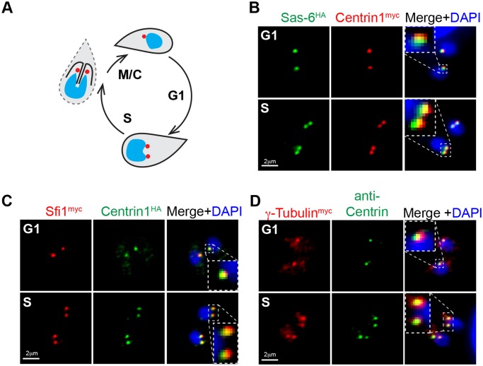
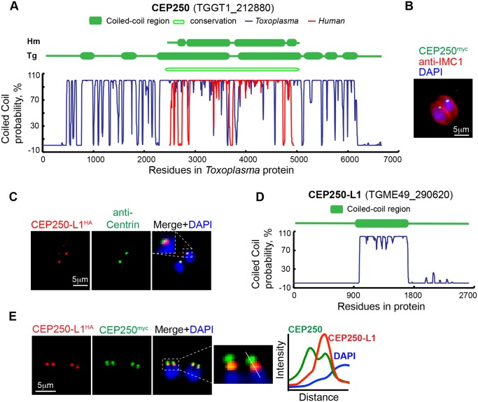
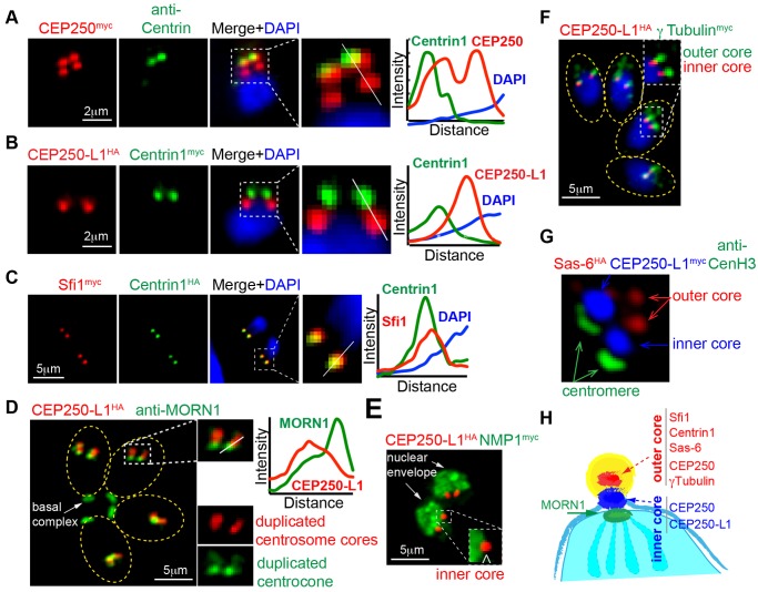
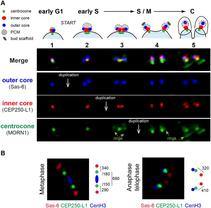
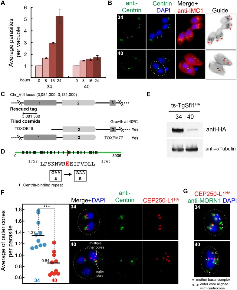
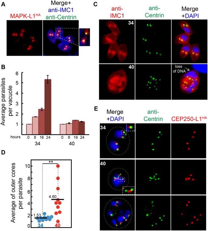
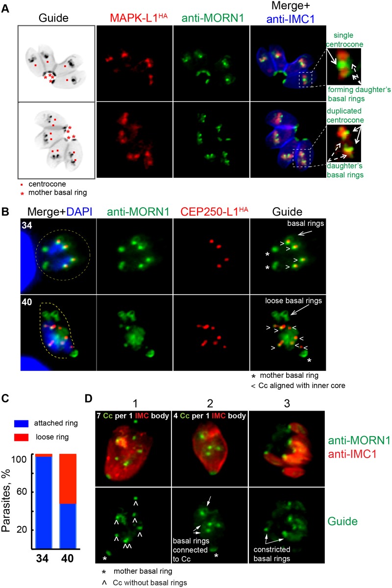
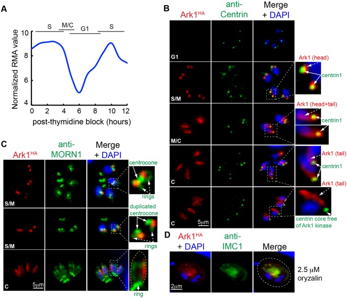
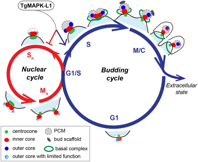
Apicomplexan parasites can change fundamental features of cell division during their life cycles, suspending cytokinesis when needed and changing proliferative scale in different hosts and tissues. The structural and molecular basis for this remarkable cell cycle flexibility is not fully understood, although the centrosome serves a key role in determining when and how much replication will occur. Here we describe the discovery of multiple replicating core complexes with distinct protein composition and function in the centrosome of Toxoplasma gondii. An outer core complex distal from the nucleus contains the TgCentrin1/TgSfi1 protein pair, along with the cartwheel protein TgSas-6 and a novel Aurora-related kinase, while an inner core closely aligned with the unique spindle pole (centrocone) holds distant orthologs of the CEP250/C-Nap protein family. This outer/inner spatial relationship of centrosome cores is maintained throughout the cell cycle. When in metaphase, the duplicated cores align to opposite sides of the kinetochores in a linear array. As parasites transition into S phase, the cores sequentially duplicate, outer core first and inner core second, ensuring that each daughter parasite inherits one copy of each type of centrosome core. A key serine/threonine kinase distantly related to the MAPK family is localized to the centrosome, where it restricts core duplication to once per cycle and ensures the proper formation of new daughter parasites. Genetic analysis of the outer core in a temperature-sensitive mutant demonstrated this core functions primarily in cytokinesis. An inhibition of ts-TgSfi1 function at high temperature caused the loss of outer cores and a severe block to budding, while at the same time the inner core amplified along with the unique spindle pole, indicating the inner core and spindle pole are independent and co-regulated. The discovery of a novel bipartite organization in the parasite centrosome that segregates the functions of karyokinesis and cytokinesis provides an explanation for how cell cycle flexibility is achieved in apicomplexan life cycles.


Conflict of interest statement

The authors have declared that no competing interests exist.

Figures

Fig 1
Fig 1. Conserved centrosomal proteins co-localize with TgCentrin1 in tachyzoites.
(A) Schematic representation of the T. gondii tachyzoite centrosome cycle. Centrosome duplication occurs at the G1/S transition, and at the completion of mitosis and budding (M/C) each new daughter parasite inherits a single TgCentrin1-containing centrosome, indicated by red dot. (B) A transgenic strain expressing epitope-tagged TgSas-6HA and TgCentrin1myc produced by sequential genetic knock-in shows tight co-localization of these centrosome proteins. A single TgSas-6HA/TgCentrin1myc structure in G1 phase duplicated in parasites that had entered S phase. Cell cycle phases of individual parasites and vacuoles were determined based on well-established nuclear and cell morphological criteria [4]. (C and D) Using a similar knock-in strategy, T. gondii orthologs of Sfi1 and γ-Tubulin were epitope-tagged with 3xmyc in the genomic locus. Localization of these proteins in the centrosome was established by co-staining the γ-Tubulinmyc transgenic strain with anti-Centrin antibody, and in the TgSfi1myc transgenic clone, epitope-tagging of TgCentrin1 with 3xHA by genetic knock-in was used. DAPI co-staining (blue) was used to visualize nucleus. Magnified inset images inside the merged images highlight the tight co-localization of the each pair of centrosomal factors in a defined centrosome core structure.
Fig 2
Fig 2. T. gondii CEP250-related proteins localize to the tachyzoite centrosome.
(A) Assessed by primary and secondary protein analysis, TgCEP250 was designated the ortholog of hCEP250. Coiled-coil domains exceeding 200 amino acids are shown in the diagram above the plots of the Marcoil prediction (http://toolkit.tuebingen.mpg.de/marcoil). Variation of the coiled-coil probability for the full length TgCEP250 protein is shown with blue line and for hCEP250 protein with red line. Primary sequence homology was determined by pBLAST and is indicated as an open block in the diagram (conservation). (B) C-terminal epitope-tagging of TgCEP250 (green) protein with 3xmyc by genetic knock-in at the chromosome locus revealed that this factor is localized to a small perinuclear structure consistent with location of the centrosome. Co-staining with DAPI (blue) and anti-IMC1 antibody (red) was used to reference nucleus and the parasite cytoskeleton, respectively. (C) The novel TgCEP250-like protein 1 (TgCEP250-L1) tagged with 3xHA-epitope (genetic knock-in) co-localized near TgCentrin1-positive structures (co-staining with anti-Centrin antibody). Inset panel shows close perinuclear localization of the merged markers. (D) The TgCEP250-L1 protein has a single predicted coiled-coil domain (http://toolkit.tuebingen.mpg.de/marcoil). (E) Co-staining of TgCEP250myc (green) and TgCEP250L-1HA (red) proteins in a transgenic clone, in which the factors were sequentially epitope-tagged by genetic knock-in, shows TgCEP250myc stains four foci in mitotic parasites, while TgCEP250-L1HA co-localized only in the two foci closest to the nucleus. Quantitative fluorescence density plot was built using data taken from a cross section of the image indicated by the white line in the enlarged merged image immediately to the left of the graph.
Fig 3
Fig 3. The tachyzoite centrosome has a unique bipartite internal organization.
The pairwise localization of several centrosome proteins was examined in tachyzoites undergoing mitosis. Detection of each protein pair was accomplished by epitope-tagging (genetic knock-in) or by using antibody raised against recombinant protein. These experiments revealed two distinct protein core complexes are contained within the tachyzoite centrosome that duplicate during the cell cycle. Two pairwise protein IFA analyses highlight these structures; the (A) upper panel is co-staining of epitope-tagged TgCEP250myc with anti-myc (red) and anti-Centrin (green). Note that TgCEP250myc is localized to the TgCentrin1 core structure (designated the outer core) as well as a novel second core (inner core) that was closer to the parasite nucleus in all cell cycle phases. The (B) panel is a dual epitope-tagged strain TgCEP250-L1HA/TgCentrin1myc co-stained with anti-HA (red) and anti-myc (green). The CEP250-related protein, TgCEP250L-1 is preferentially localized to the novel inner core that lacks TgCentrin1. (C) A recognized co-factor of TgCentrin1, TgSfi1 (red), exclusively co-localized with TgCentrin1 (green) in the outer centrosomal core (co-staining of the dual-tagged clone TgCentrin1HA/TgSfi1myc). Differential composition of the outer versus inner centrosome core also included outer core proteins TgSas-6 and Aurora-related kinase 1 (TgARK1) described below. (D) Duplicated centrosomal cores containing TgCEP250-L1HA were segregated from the centrocone structure embedded in the nuclear envelope as demonstrated by co-staining with anti-TgMORN1 antibody (green). (E) The separation of the inner core from the nuclear envelope was further established by co-staining for the nuclear mesh protein, TgNMP1; analysis of dual-tagged clone TgCEP250-L1HA/TgNMP1myc. Note that inner core containing TgCEP250-L1HA is localized to the cytoplasmic interface of the nucleus in the inset image. (F) T. gondii γ-Tubulin is localized to the TgCentrin1-associated outer core (see Fig. 1D) that is distal from the inner core co-stained for TgCEP250-L1HA; shown here is dual-tagged clone TgCEP250-L1HA/γ-Tubulinmyc. (G) The spatial segregation of the bipartite core structures of the Toxoplasma centrosome is well resolved and aligned with the centromeres as captured by super-resolution microscopy; markers used were outer core = TgSas-6HA, inner core = TgCEP250-L1myc, and the anti-TgCenH3 antibody to detect the centromere/kinetochore intranuclear structure [25]. For image panels A-D, quantitative fluorescence density plots were included. The data for each density graph was taken from a cross section of the image indicated by the white line in the enlarged merged image immediately to the left of the graph. (H) Diagram is included that summarizes the discovery of two replicating centrosome cores (one-half of the mitotic organization) with differential protein composition.
Fig 4
Fig 4. Morphogenesis of the centrosome during the tachyzoite cell cycle.
(A) Morphological transitions of the outer and inner protein core complexes were monitored in the dual epitope-tagged TgSas-6HA/TgCEP250-L1myc clone. Co-staining with antibodies against TgMORN1 was used to visualize the nuclear membrane centrocone compartment (spindle pole). Above the fluorescent merged images (Merge) a diagram of the composite structures is placed within the cell cycle timing of the biosynthetic events. Five distinctive morphological transitions were identified, and three sequential duplication events (white arrows) were recorded in the following order: duplication of the outer core (TgSas-6HA, blue panel) followed by the inner core (TgCEP250-L1myc, red panel) and lastly, the centrocone (anti-TgMORN1 antibody, green panel). The cell cycle stages for each transition of the centrosome cycle were determined using well-established nuclear and cellular morphologies. (B) Super resolution images of the centrosome cores (TgSas-6HA/TgCEP250-L1myc) in relation to the centromeres (visualized with anti-TgCenH3 antibody) in the tachyzoite metaphase and anaphase/telophase. The diagram to the right of each image estimates the physical position and distance between the middle points of the three mitotic structures: outer and inner centrosome cores and kinetochore.
Fig 5
Fig 5. The outer centrosome core protein TgSfi1 has an essential role in the parasite cell cycle.
(A) Growth of the chemical mutant 9–86E4 is inhibited by high temperature, leading to lethal arrest (40°C). Cultures were pre-synchronized by limited invasion, and the average number of parasites per vacuole was calculated after 0, 8, 16, and 24 h at indicated temperature. Bar graph shows mean values and standard deviations for three growth experiments with a minimum of 50 vacuoles per time point (for all raw data, see S1 Data). (B) Duplication of the outer core detected by anti-Centrin staining is severely affected in the 9–86E4 mutant grown at 40°C for 20 hours but not when the mutant is grown at the permissive temperature (34°C). Culture temperatures are indicated in the upper left of each image panel. The included guide panel is a non-colored inverted image of the merged red (anti-IMC1) and green (anti-Centrin) stains. Red asterisks indicate the position of the Centrin-positive structures. Note the parasite at 40°C with duplicated nuclei with a single TgCentrin1-associated core (circled) where, normally, there should be two cores. (C) Genetic complementation of mutant 9–86E4 with cosmid genomic libraries identified the defective locus on chromosome VIII (see top diagram) and this was further resolved to gene 2 (TGME49_274000) with individual cosmids that span the locus; gene 2 encodes centrin co-factor, TgSfi1 (see S1B Fig. and S2 Fig.) [38]. (D) Whole genome sequencing of mutant 9–86E4 independently confirmed the ts-TgSfi1 protein was mutated with a E1759K change shown by a red bar in the TgSfi1 protein diagram. Putative centrin binding sites (black box) are also indicated (see also S2 Fig.). (E) Western blot analysis of the mutant 9–86E4 parasites expressing ts-TgSfi1HA protein after 24 h growth at 34°C or 40°C. Total lysate of 20 x 106 parasites were probed with anti-HA-epitope or anti-α-Tubulin antibodies. (F) The average of TgCentrin1 containing outer cores (anti-Centrin staining) per parasite, determined by anti-IMC1 staining of the mother parasite, was quantified in ten microscopic fields with an average of 3–10 vacuoles (for all raw data, see S1 Data) revealing a significant reduction of the outer cores when mutant 9–86E4 was shifted to 40°C (red dots). P-value ≤ 0.0005 (***) was calculated using paired two-tail t test. The loss of the outer core at high temperature in the ts-TgSfi1 mutant does not prevent replication of the inner core. To monitor inner core, TgCEP250-L1HA was introduced in the mutant 9–86E4 (see Generation of transgenic tachyzoite strains in Materials and Methods). Co-staining of TgCEP250-L1HA (inner core) and anti-Centrin (outer core) showed that amplification of the inner core occurred in parasites in which duplication of the outer core was inhibited. (G) Multiple inner cores (TgCEP250-L1HA, red) in mutant 9–86E4 parasites at 40°C showed tight alignment and matched duplication of the nuclear centrocone (anti-TgMORN1stain, green).
Fig 6
Fig 6. TgMAPK-L1 localizes to the centrosome and controls proper duplication.
(A) Co-staining of the endogenously tagged TgMAPK-L1HA with anti-Centrin and anti-IMC1 antibodies. Magnified image on the right shows a vacuole of four parasites post-duplication of the centrosome surrounded by TgMAPK-L1HA in the pericentrosomal matrix (PCM). (B) Mutant 11–31G12 parasites defective in TgMAPK-L1 (see details in S4 Fig.) quickly growth arrested at 40°C. Growth of the mutant at 34°C and 40°C was quantified as described in the Fig. 5A (for all raw data, see S1 Data). (C) Disruption of ts-TgMAPK-L1 function resulted in the over-duplication of the outer centrosomal cores (anti-Centrin, green) accompanied by defects in chromosome segregation and nuclear division (double-headed arrow). (D) Quantification of the Centrin-containing outer cores per original mother parasite (determined by anti-IMC1 staining) in mutant 11–31G12 parasites grown at 34°C (blue dots) and 40°C (red dots) for 20 h is shown in the dot plot (for all raw data see S1 Data). Mean values for each dataset are also indicated (black bars). Significant difference between two conditions was verified by paired t test that returned the p-value ≤ 0.007 (**). Note that while both cores are amplified beyond the normal stoichiometry at 40°C, there is not a precise match in the total number of outer and inner cores in every parasite. (E) To determine the fate of the inner centrosomal core, the TgCEP250-L1 protein epitope-tagged with 3xHA (red) was introduced into mutant 11–31G12 parasites, producing a transgenic clone, 11–31G12(TgCEP250L-1HA). Co-staining of this clone with anti-HA (red) and anti-Centrin antibodies (green) showed that at 40°C the inner cores were multiplied along with the outer centrosome cores while preserving proper spatial alignment. Duplication of the inner core was slightly delayed, leading to accumulation of the intermediate “dumbbell” structures shown in the inset of the middle 40°C merged panel. Yellow dotted line indicates vacuole boundary in the infected host cell shown in these images.
Fig 7
Fig 7. TgMAPK-L1 has a key role in coupling mitosis and cytokinesis.
(A) In replicating parasites endogenously tagged wild-type TgMAPK-L1HA (red stain) showed a predominant alignment with single (upper panel) and duplicated (lower panel) centrocone (Cc; anti-MORN1 staining), surrounded by the TgMORN1-rings (new daughter’s basal complexes). Non-color guide panels of the inverted merged images of the anti-IMC1 (blue channel) and the anti-MORN1 (green channel) staining were included here to provide a reference to the location and scale of the subcellular structures observed within the parasite. Magnified merged images to the right of each panel are included to provide greater detail of the key structures stained. (B) Amplified inner cores (TgCEP250-L1HA) in ts-TgMAPK-L1 mutant 11–31G12 parasites arrested at 40°C for 20 h maintained alignment with the nuclear centrocone (anti-TgMORN1 staining); however, MORN1-rings (daughter’s basal rings) were no longer attached to duplicated centrocones in many parasites. Asterisks indicate position of the basal complex of the mother cell and “<” points to the aligned inner core/centrocone structures. Examples of disconnected basal rings are evident in the lower 40°C panel (long arrow), while at the permissive temperature (34°C) the daughter’s basal rings are tightly connected to the centrocone. Yellow dashed line in the merged images indicates the parasite size (40) or the size of the vacuole (34). (C) Bar graph shows quantification of the disruption of the daughter’s MORN1-ring attachment to the centrocone. Only S/M stages that normally display MORN1-rings were quantified at 34°C. Vacuoles with detectable MORN1-rings were analyzed at 40°C. No less than 100 vacuoles were analyzed for each condition (for all raw data see S1 Data). (D) Co-staining of the nuclear centrocone and basal rings (anti-TgMORN1 antibody, green) with the membrane scaffold (anti-IMC1 antibody) in the ts-TgMAPK-L1 parasites arrested at 40°C for 20 h. Merged images are shown in the upper panel. Anti-TgMORN1 stain in the Guide panel highlights only the centrocone and basal ring relationships. The three images are representative of the predominant morphological defects associated with ts-TgMAPK-L1 in mutant parasites. First image represents an early step in the development of phenotype: mother cell with multiplied centrocones (7) and no visible formation of the daughter’s basal rings (“^”). The mother cell in the second image similarly has amplified centrocones (4), however, they are connected to the well-developed daughter MORN-rings (arrows). The consecutive step of abnormal multiple budding (7) is represented on the third image. Note that each daughter cell has a constricted basal ring (arrow) at the proximal end of the body.
Fig 8
Fig 8. A novel Toxoplasma aurora-related kinase shows dynamic association with parasite cytoskeletal structures.
Periodicity of TgArk1 mRNA (A) translates into dynamic cell cycle protein expression (B) (for raw mRNA data, see S1 Data). At peak expression in S/M phase, TgArk1HA protein (red, 3xHA tagged in genome by knock-in) was co-localized to the centrosome outer core complex as demonstrated by the anti-HA (red) and anti-Centrin (green) co-staining. Intriguingly, as parasites entered mitosis, TgArk1HA also became localized to a discrete linear structure. The dual pattern of localization in the centrosome outer core (TgArk1, head morphology) and the novel linear structure (TgArk1, tail morphology) was altered further in late cytokinesis (last two panels) when TgArk1 disappeared from the TgCentrin1-associated outer core complex. Magnified images of the specific areas within the merged panels are shown to the right. Cell cycle stages are indicated in the upper left of each image series. (C) Co-staining with anti-TgMORN1 (green) antibody showed that association of TgArk1HA with the centrosome is maintained during centrocone development and duplication. The linear structure stained by TgArk1HA (tail morphology) is unusual and runs along one side of the growing daughter bud. Yellow dotted line in the magnified image of the cytokinesis stage represents a cytoskeleton of the growing daughter bud. Cell cycle stages are indicated in the upper left of each image series. (D) Treatment of tachyzoites with 2.5 μM oryzalin for 24 h to disrupt subpellicular microtubules indicates the linear TgArk1HA-tail is an unknown cytoskeletal structure, although the primary association may also involve the factors of the inner membrane complex because TgArk1HA staining was retained with the disrupted IMC1-containing material after oryzalin caused disassembly of the daughter subpellicular microtubules. Dotted line outlines boundary of the parasitophorous vacuole in the infected host cell.
Fig 9
Fig 9. Regulation of the apicomplexan centrosome cycle may be key to cell division flexibility.
The two basic cell cycles responsible for Apicomplexa replication are diagrammed with the proposed centrosome cycles overlaid. Entering the single budding cycle produces daughter parasites, while multiple nuclear cycles amplify chromosome number in the absence of budding. In this model, modulation of the complex centrosome is proposed to regulate the outcome of each cell division cycle. Active mechanisms associated with the outer centrosome core (bright blue circle) control the initiation of budding (green open ring, basal complex; gray oval, bud scaffold), while inactivation of this core (light blue circle) may be required to suspend budding in the nuclear cycle. In the absence of an active outer core, the inner core and centrocone permit each nucleus to complete chromosome replication and mitosis. Our study points to a key PCM factor, TgMAPK-L1, as a regulator of counting in the budding cycle of tachyzoites. Operating at the G1/S boundary, we propose that TgMAPK-L1 may promote and restrict the biosynthetic outcome of the budding cycle (blue arrow), while blocking a path into the nuclear cycle (red arrow).

Comment in

References

    1. Arnot DE, Ronander E, Bengtsson DC (2011) The progression of the intra-erythrocytic cell cycle of Plasmodium falciparum and the role of the centriolar plaques in asynchronous mitotic division during schizogony. Int J Parasitol 41: 71–80. 10.1016/j.ijpara.2010.07.012 - DOI - PubMed
    1. Sogin ML, Silberman JD (1998) Evolution of the protists and protistan parasites from the perspective of molecular systematics. Int J Parasitol 28: 11–20. - PubMed
    1. Anderson-White B, Beck JR, Chen CT, Meissner M, Bradley PJ, et al. (2012) Cytoskeleton assembly in Toxoplasma gondii cell division. International review of cell and molecular biology 298: 1–31. 10.1016/B978-0-12-394309-5.00001-8 - DOI - PMC - PubMed
    1. Francia ME, Striepen B (2014) Cell division in apicomplexan parasites. Nat Rev Microbiol 12: 125–136. 10.1038/nrmicro3184 - DOI - PubMed
    1. White MW, Radke JR, Radke JB (2014) Toxoplasma development—turn the switch on or off? Cell Microbiol 16: 466–472. 10.1111/cmi.12267 - DOI - PubMed

Publication types

MeSH terms