. 2015;9(4):3530-9.
doi: 10.1021/acsnano.5b00161. Epub 2015 Mar 19.

Membrane-assisted growth of DNA origami nanostructure arrays

Affiliations

Membrane-assisted growth of DNA origami nanostructure arrays

Samet Kocabey et al. ACS Nano. 2015.

Abstract

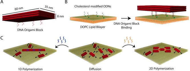
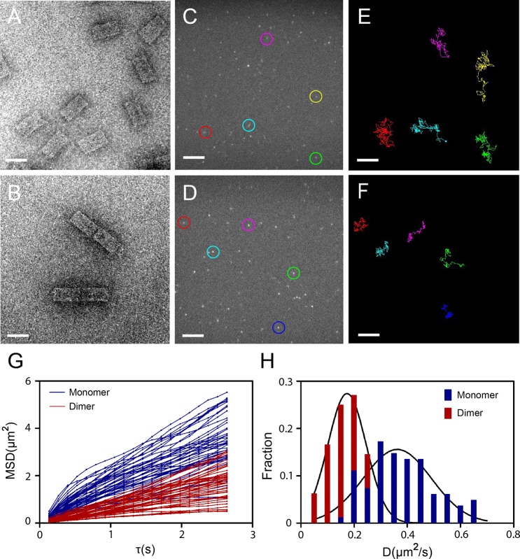
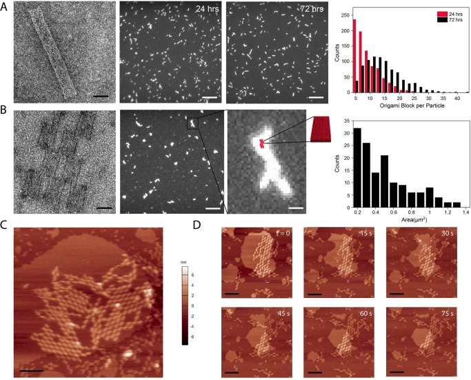
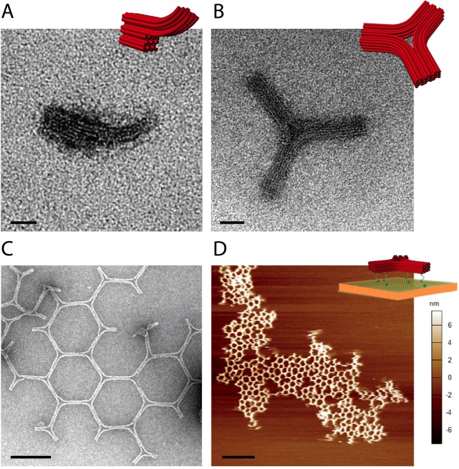
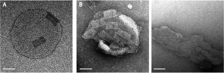
Biological membranes fulfill many important tasks within living organisms. In addition to separating cellular volumes, membranes confine the space available to membrane-associated proteins to two dimensions (2D), which greatly increases their probability to interact with each other and assemble into multiprotein complexes. We here employed two DNA origami structures functionalized with cholesterol moieties as membrane anchors--a three-layered rectangular block and a Y-shaped DNA structure--to mimic membrane-assisted assembly into hierarchical superstructures on supported lipid bilayers and small unilamellar vesicles. As designed, the DNA constructs adhered to the lipid bilayers mediated by the cholesterol anchors and diffused freely in 2D with diffusion coefficients depending on their size and number of cholesterol modifications. Different sets of multimerization oligonucleotides added to bilayer-bound origami block structures induced the growth of either linear polymers or two-dimensional lattices on the membrane. Y-shaped DNA origami structures associated into triskelion homotrimers and further assembled into weakly ordered arrays of hexagons and pentagons, which resembled the geometry of clathrin-coated pits. Our results demonstrate the potential to realize artificial self-assembling systems that mimic the hierarchical formation of polyhedral lattices on cytoplasmic membranes.

Keywords: DNA nanotechnology; DNA origami; arrays; cholesterol; clathrin; diffusion; lipid membrane.


Figures

Figure 1
Figure 1
DNA origami block on lipid membrane. (A) DNA origami block structure consisting of three layers of 14 double helices each. The indicated dimensions assume a distance between the base pairs of 0.34 nm and an average distance between the centers of the helices of 2.5 nm. (B) Cholesterol-mediated binding of origami blocks to a lipid bilayer membrane (ODN: oligodeoxynucleotide; DOPC: 1,2-dioleoyl-sn-glycero-3-phosphocholine). (C) Programmed polymerization of DNA origami blocks into different superstructures following the addition of connector staples to structures diffusing on the membrane.
Figure 2
Figure 2
Lateral diffusion of origami block monomers and dimers on supported lipid bilayers. TEM images of DNA origami block monomers (A) and dimers (B) (scale bars: 50 nm). Fluorescence images of DNA origami block monomers (C) and dimers (D) on a DOPC lipid bilayer (scale bars: 5 μm). Example diffusion trajectories for five block monomers (E) and five dimers (F) (scale bars: 5 μm). (G) Time-dependent mean-square displacement (MSD) plot of monomers and dimers. (H) Distribution of diffusion coefficients obtained from single-particle tracking of origami block monomers and dimers. The black lines are Gaussian fits to the distributions.
Figure 3
Figure 3
Programmable polymerization of DNA origami blocks on supported lipid bilayers. (A) One-dimensional polymerization. Left: TEM image of polymerized origami blocks after 24 h of incubation in TE buffer with 11 mM Mg2+ (scale bar: 50 nm). Middle: Fluorescence images of polymerized origami blocks after 24 and 72 h of incubation on DOPC lipid bilayers (scale bar: 5 μm). Right: Histogram of the estimated number of origami blocks per fluorescent particle after 24 and 72 h of incubation. (B) Two-dimensional polymerization. Left: TEM image of polymerized DNA origami blocks after 24 h of incubation in TE buffer with 11 mM Mg2+ (scale bar: 40 nm). Middle: Fluorescence image of polymerized DNA origami blocks after 72 h of incubation (scale bar: 5 μm). Inset: Magnified image of one of the particles including a scheme of a 2D origami lattice for size comparison (scale bar: 400 nm). Right: Histogram of the area of the lattices after 72 h of incubation. (C) AFM image demonstrating lattice formation on the lipid bilayer (scale bar: 300 nm, scan rate: 10 Hz, 512 × 512 pixels). (D) AFM images showing the decomposition of a lipid bilayer over a time interval of 75 s, which results in the adsorption of an origami lattice on the mica surface (scale bar: 300 nm, scan rate: 10 Hz, 1024 × 1024 pixels).
Figure 4
Figure 4
Programmable polymerization of DNA origami triskelions. (A) TEM image of a truncated Y-shaped DNA origami (scale bar: 20 nm). (B) TEM image of a triskelion DNA origami assembled from three truncated Y structures (scale bar: 20 nm). (C) TEM image showing the polymerization of DNA origami triskelions into hexagonal lattices in solution (scale bar: 200 nm). (D) AFM image demonstrating extended polymerization of DNA origami trimers into 2D arrays on supported lipid bilayer (scale bar: 2 μm, scan rate: 4 Hz, 1024 × 1024 pixels).
Figure 5
Figure 5
DNA origami block polymerization on SUVs. (A) TEM image of DNA origami block monomers on an SUV (scale bar: 60 nm). (B) 2D lattice formation of the DNA origami blocks on SUVs (scale bars: 60 nm). Interactions between origami lattices and SUVs apparently result in a shape deformation or even destruction of the vesicles.

Comment in

References

    1. Axelrod D. Lateral Motion of Membrane Proteins and Biological Function. J. Membr. Biol. 1983, 75, 1–10. - PubMed
    1. Heldin C. H. Dimerization of Cell Surface Receptors in Signal Transduction. Cell 1995, 80, 213–223. - PubMed
    1. Lamb T. D. Gain and Kinetics of Activation in the G-Protein Cascade of Phototransduction. Proc. Natl. Acad. Sci. U.S.A. 1996, 93, 566–570. - PMC - PubMed
    1. Pearse B. M. Clathrin: A Unique Protein Associated with Intracellular Transfer of Membrane by Coated Vesicles. Proc. Natl. Acad. Sci. U.S.A. 1976, 73, 1255–1259. - PMC - PubMed
    1. Doherty G. J.; McMahon H. T. Mechanisms of Endocytosis. Annu. Rev. Biochem. 2009, 78, 857–902. - PubMed

Publication types