. 2015 Apr;20(4):433-43.
doi: 10.1007/s10495-015-1091-7.

Exposure to the complement C5b-9 complex sensitizes 661W photoreceptor cells to both apoptosis and necroptosis

Affiliations

Exposure to the complement C5b-9 complex sensitizes 661W photoreceptor cells to both apoptosis and necroptosis

Hui Shi et al. Apoptosis. 2015 Apr.

Abstract

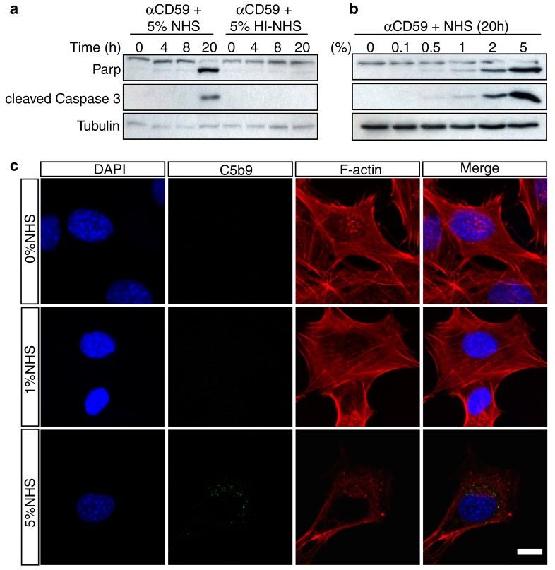
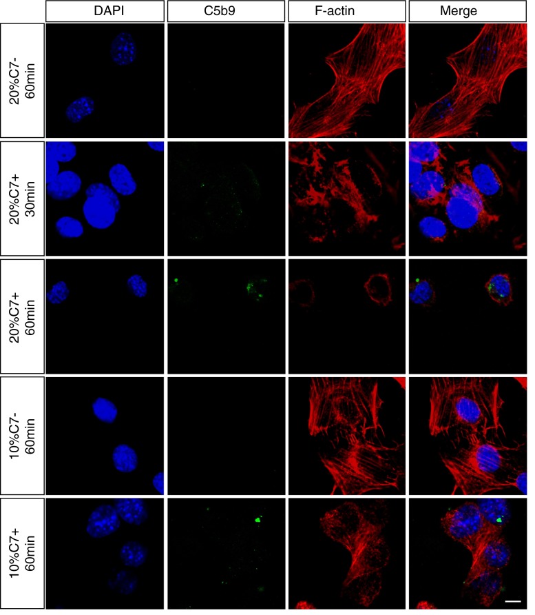
The loss of photoreceptors is the defining characteristic of many retinal degenerative diseases, but the mechanisms that regulate photoreceptor cell death are not fully understood. Here we have used the 661W cone photoreceptor cell line to ask whether exposure to the terminal complement complex C5b-9 induces cell death and/or modulates the sensitivity of these cells to other cellular stressors. 661W cone photoreceptors were exposed to complete normal human serum following antibody blockade of CD59. Apoptosis induction was assessed morphologically, by flow cytometry, and on western blotting by probing for cleaved PARP and activated caspase-3. Necroptosis was assessed by flow cytometry and Sirtuin 2 inhibition using 2-cyano-3-[5-(2,5-dichlorophenyl)-2-furyl]-N-5-quinolinylacrylamide (AGK2). The sensitivity of 661W cells to ionomycin, staurosporine, peroxide and chelerythrine was also investigated, with or without prior formation of C5b-9. 661W cells underwent apoptotic cell death following exposure to C5b-9, as judged by poly(ADP-ribose) polymerase 1 cleavage and activation of caspase-3. We also observed apoptotic cell death in response to staurosporine, but 661W cells were resistant to both ionomycin and peroxide. Interestingly, C5b-9 significantly increased 661W sensitivity to staurosporine-induced apoptosis and necroptosis. These studies show that low levels of C5b-9 on 661W cells can induce apoptosis, and that C5b-9 specifically sensitizes 661W cells to certain apoptotic and necroptotic pathways. Our observations provide new insight into the potential role of the complement system in photoreceptor loss, with implications for the molecular aetiology of retinal disease.


Figures

Fig. 1
Fig. 1
Validation of the 661W cell line. a We used mouse and rat genomic DNA as templates to confirm the species of the cell line, with mouse ß-globin and rat ß-actin probes respectively. Lane 1 661W cell genomic DNA; Lane 2 rat genomic DNA; Lane 3 mouse genomic DNA; Lane 4 water (negative control). b We then used a set of mouse-specific retinal primers as indicated in the figure to assess expression of rod and cone markers in the cell line. Lane 1 661W cell cDNA; Lane 2 mouse neuroretina cDNA (positive control); Lane 3 mouse liver cDNA (negative control); Lane 4 water (negative control)
Fig. 2
Fig. 2
Formation of C5b-9 on 661W cells. a MTT viability assay measuring the extent of 661W cell death following treatment with blocking antibodies to CD59 and increasing concentrations (5, 15, 25, 30 %) of NHS for 1 h at 37 °C. For the negative control, cells were treated with anti-CD59 and 30 % HI-NHS, and for the positive control with 1 % Triton X-100. Data are plotted as mean ± S.D. (n = 3). b 661W cells were immunostained for C5b-9 and counterstained with DAPI to visualise nuclei. Cells were treated with 5 % NHS alone, anti-CD59+ 5 % NHS, 10 % NHS alone, or anti-CD59+ 10 % NHS. C5b-9 was barely visible at the lower serum concentration or in cells that weren’t treated with the CD59 blocking antibody, but appeared in punctate form in cells exposed to 10 % NHS (zoomed image). Scale bar = 10 μm. All experiments were repeated at least three times, and representative images are shown
Fig. 3
Fig. 3
C5b-9 induces signature features of apoptosis in 661W cells in a dose- and time-dependent manner. a 661W cells were serum-starved for 24 h, then treated with anti-CD59 for 1 h followed by 5 % NHS for 1 h. Cells were lysed after 0, 4, 8 and 20 h and subjected to immunoblot analysis for PARP and cleaved caspase 3. Cells were treated with 5 % HI-NHS as a negative control and α-tubulin was included as a loading control. The blot shows that cleavage of PARP and activation of caspase-3 after 20 h occurred only in cells treated with NHS. b 661W cells were prepared as in (a), and treated with different concentrations of NHS for 20 h before immunoblotting. PARP cleavage and caspase-3 activation were faintly evident in 1 % NHS, but strikingly so in 2 and 5 % NHS. c 661W cells were prepared as in (a) with exposure to NHS for 20 h, then fixed, immunostained for C5b-9 and F-actin with nuclear counterstaining, and examined by confocal microscopy to investigate morphological changes. The images show that in 0 and 1 % NHS, 661W cells maintained a normal healthy morphology with abundant F-actin stress fibres, whereas in 5 % NHS surviving cells typically were smaller with fewer stress fibres and F-actin clumping. Scale bar 10 μm. All experiments were repeated at least three times, and representative blots/images are shown
Fig. 4
Fig. 4
C7 is required for C5b-9 formation. Serum-deprived and anti-CD59 treated 661W cells were treated with either C7-depleted NHS (10 or 20 % for 60 min) or C7-depleted serum supplemented with purified exogenous C7 (100 µg/ml) (10 or 20 % for 60 min). Immunofluorescence staining and confocal imaging of C5b-9 and F-actin shows that in cells treated with C7 deficient serum, cellular morphology was normal with abundant F-actin stress fibres, and no evidence of C5b-9 staining. Restoration of C7 led to the appearance of punctate C5b-9 staining, and characteristic disruption of the F-actin cytoskeleton. DAPI was used to stain the nuclei. Scale bar 10 μm. All experiments were repeated at least three times, and representative images are shown
Fig. 5
Fig. 5
C5b-9 sensitizes 661W cells to staurosporine-mediated apoptosis. Cell apoptosis was detected by immunoblotting for PARP cleavage and activated caspase 3. a Serum-deprived and anti-CD59 treated 661W cells were incubated with increasing concentrations of NHS (0, 0.1, 0.5, 1, 2, 5 %) for 1 h, then with complete medium alone or with 100nM staurosporine (SS), 1 µM ionomycin (INM) or 10 µM H2O2 for 8 h. b 661W apoptosis was compared between cells treated with 100 nM staurosporine or medium alone for 0, 4, 8 and 20 h, which revealed PARP cleavage and caspase-3 activation only at 20 h. The kinetics of staurosporine-induced apoptosis were then investigated following treatment of 661W cells with either 5 % NHS or 5 % HI-NHS for 1 h, which revealed PARP cleavage and caspase-3 activation at 8 h only in the presence of 5 % NHS. c 661W cells were treated with a range of concentrations of chelerythrine for 4, 8 and 12 h. The immunoblots show rapid onset of PARP cleavage and caspase-3 activation with 10 and 20 µM agonist. d The sensitivity of 661W cells to chelerythrine was examined with and without prior exposure to 5 % NHS or 5 % HI-NHS. The immunoblots show that at 4 and 8 h, there was no difference between 5 % NHS and 5 % HI-NHS, though in both conditions PARP cleavage and caspase-3 activation were enhanced compared to chelerythrine alone (c)
Fig. 6
Fig. 6
Chelerythrine and staurosporine induce apoptosis and necroptosis in 661W cells. Cell death was examined by Annexin V/PI staining and flow cytometry. a Quantitation of apoptotic 661W cells following treatment with a range of concentrations of staurosporine (0–100 nM) for 4 h and 8 h in the presence of 5 % NHS or 5 % HI-NHS. b Example of the primary data for the 8 h, 100 nM staurosporine experiment. Apoptotic cells are Annexin V+ and locate to the top-left quadrant of the scatter plot whereas necroptotic cells are Annexin V and PI double positive and locate to the upper right quadrant. c Quantitation of necroptotic 661W cells from the same experiment as (a) and (b). d Necroptotic 661W cells were quantified as in (c) following treatment with 100 nM staurosporine for 8 h ± 10 µM AGK2. Necroptosis was significantly inhibited by AGK2. e Serum-starved and anti-CD59 treated 661W cells were incubated with 5 % NHS or 5 % HI-NHS for 1 h and then incubated with 0–20 µM chelerythrine for 4 or 8 h. Cells were then stained and subjected to flow cytometry as described above. The bar charts show the percentages of apoptotic cells. f Quantitation of necroptotic 661W cells from the same experiment as (e). g Representative scatter plots showing the results for cells treated with 20 µM chelerythrine for 8 h. (*P < 0.05, **P < 0.01, n = 3)

References

    1. Noris M, Remuzzi G. Overview of complement activation and regulation. Semin Nephrol. 2013;33:479–492. doi: 10.1016/j.semnephrol.2013.08.001. - DOI - PMC - PubMed
    1. Ballanti E, Perricone C, Greco E, et al. Complement and autoimmunity. Immunol Res. 2013;56:477–491. doi: 10.1007/s12026-013-8422-y. - DOI - PubMed
    1. Stevens B, Allen NJ, Vazquez LE, et al. The classical complement cascade mediates CNS synapse elimination. Cell. 2007;131:1164–1178. doi: 10.1016/j.cell.2007.10.036. - DOI - PubMed
    1. Stephan AH, Barres BA, Stevens B. The complement system: an unexpected role in synaptic pruning during development and disease. Annu Rev Neurosci. 2012;35:369–389. doi: 10.1146/annurev-neuro-061010-113810. - DOI - PubMed
    1. Kim DD, Song WC. Membrane complement regulatory proteins. Clin Immunol. 2006;118:127–136. doi: 10.1016/j.clim.2005.10.014. - DOI - PubMed

Publication types