

Tubal Ligation Induces Quiescence in the Epithelia of the Fallopian Tube Fimbria
- PMID: 25736327
- PMCID: PMC5933088
- DOI: 10.1177/1933719115574345
Tubal Ligation Induces Quiescence in the Epithelia of the Fallopian Tube Fimbria
Abstract
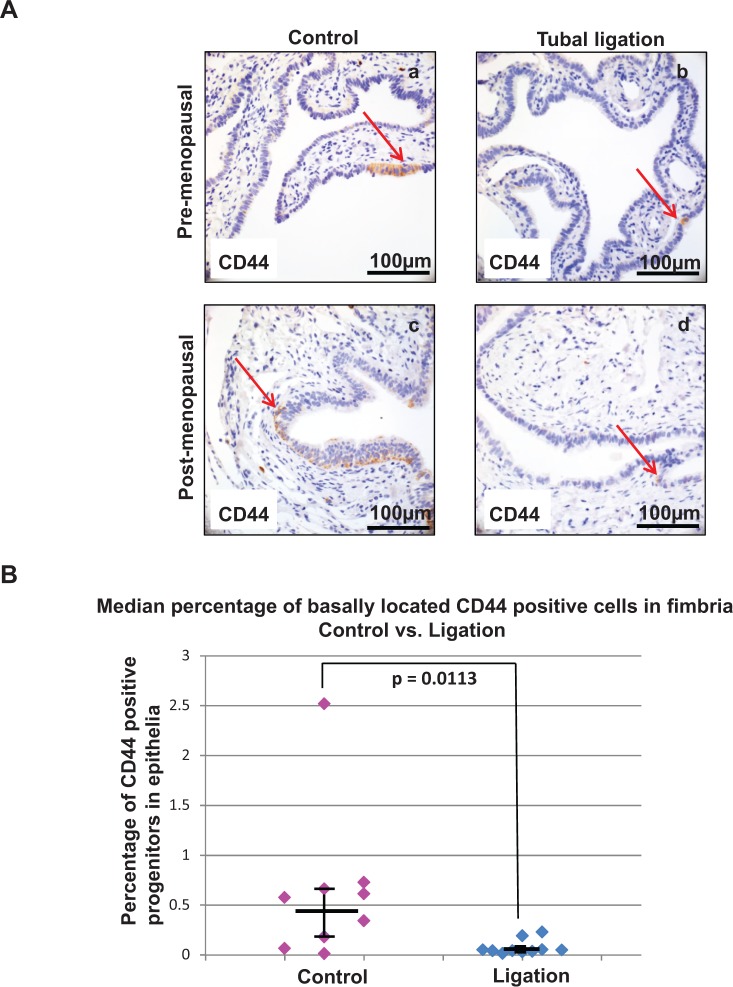
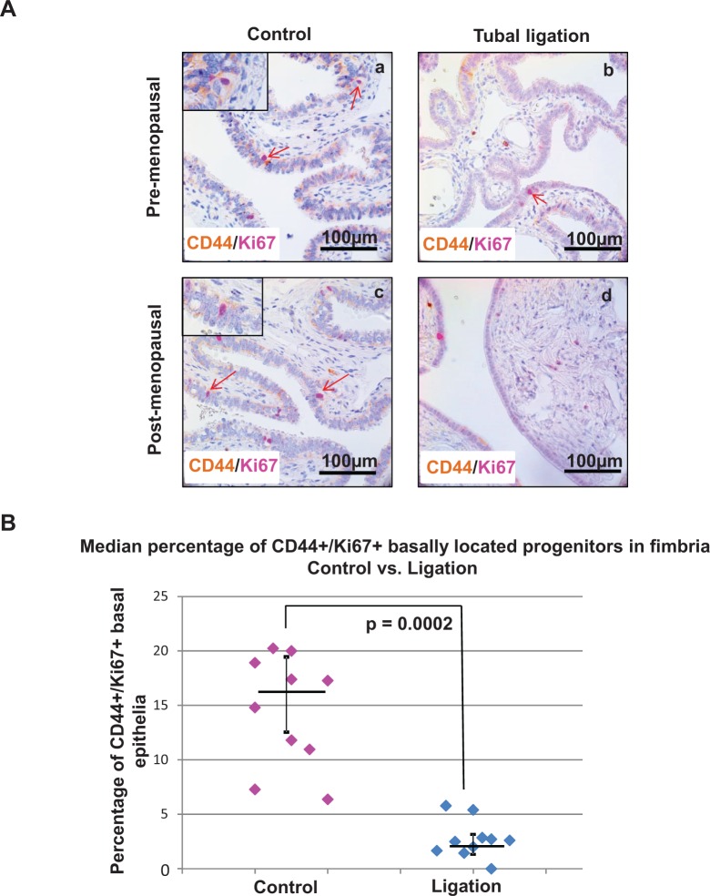
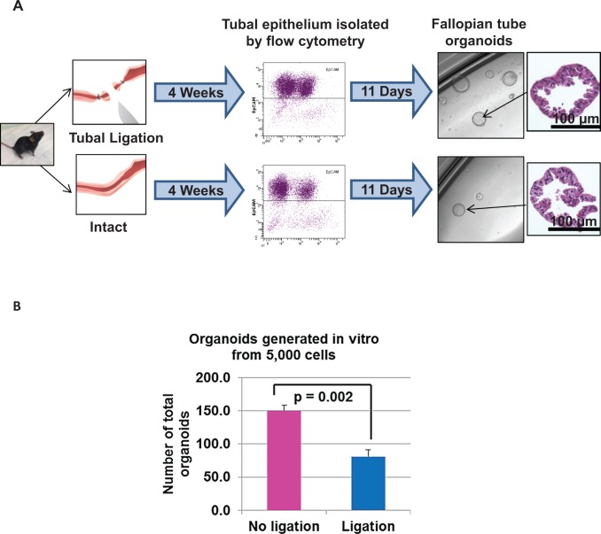
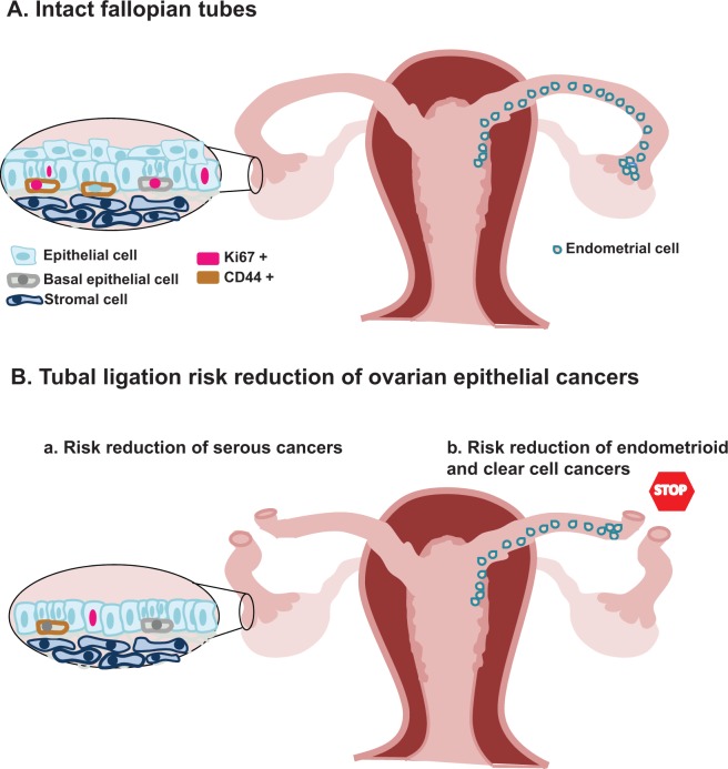
Tubal ligation keeps the fimbriated end of the fallopian tube intact while interrupting the conduit for sperm and egg between the uterus and ovary. Tubal ligation is associated with an approximately 20% decreased risk of high-grade serous ovarian cancers, which mounting evidence suggests arise from the distal fallopian tube epithelium. We postulated that biological changes at the epithelial cellular level of the distal fallopian tube may account for the surgical procedure's observed risk reduction. We compared the histology, presence of epithelial progenitors (basally located CD44-positive cells), and degree of epithelial proliferation (Ki67-positive cells) of distal fallopian tube from 10 patients with previous tubal ligation and 10 age-matched patients with uncut fallopian tubes. A significantly reduced population of proliferating epithelial progenitors (basally located CD44/Ki67 dual-positive cells) was detected in the tubal ligated specimens (P = .0002). To functionally assess the effect of tubal ligation, a murine model was utilized to compare the growth capacity of distal fallopian tube epithelial cells isolated from either ligated or sham-operated tubal epithelia. Murine fallopian tube epithelial cells isolated after tubal ligation showed a significantly reduced capacity to grow organoids in culture compared to sham-operated controls (P = .002). The findings of this study show that tubal ligation is associated with a reduced presence and decreased proliferation of progenitor cells in the distal fallopian tube epithelium. These compositional and functional changes suggest that tubal ligation induces quiescence of distal fallopian tube epithelial cells.
Keywords: fallopian epithelial progenitors; fallopian tube epithelial proliferation; tubal ligation.
© The Author(s) 2015.
Conflict of interest statement
Figures
Similar articles
-
Stem-like epithelial cells are concentrated in the distal end of the fallopian tube: a site for injury and serous cancer initiation.Stem Cells. 2012 Nov;30(11):2487-97. doi: 10.1002/stem.1207. Stem Cells. 2012. PMID: 22911892 Free PMC article.
-
Proliferation in the normal FTE is a hallmark of the follicular phase, not BRCA mutation status.Clin Cancer Res. 2012 Nov 15;18(22):6199-207. doi: 10.1158/1078-0432.CCR-12-2155. Epub 2012 Sep 11. Clin Cancer Res. 2012. PMID: 22967960
-
[Morphologic changes of fallopian tubal epithelium in ovarian serous tumors].Zhonghua Bing Li Xue Za Zhi. 2012 Jul;41(7):433-7. doi: 10.3760/cma.j.issn.0529-5807.2012.07.001. Zhonghua Bing Li Xue Za Zhi. 2012. PMID: 22932451 Chinese.
-
Fallopian tube precursors of ovarian low- and high-grade serous neoplasms.Histopathology. 2013 Jan;62(1):44-58. doi: 10.1111/his.12046. Histopathology. 2013. PMID: 23240669 Review.
-
The fallopian tube as the origin of high grade serous ovarian cancer: review of a paradigm shift.J Obstet Gynaecol Can. 2014 Feb;36(2):133-140. doi: 10.1016/S1701-2163(15)30659-9. J Obstet Gynaecol Can. 2014. PMID: 24518912 Review.
Cited by
-
Variables Affecting CA15.3 Tumor Antigen Expression and Antibodies against It in Female National Health and Nutritional Survey Participants.Cancer Epidemiol Biomarkers Prev. 2024 Sep 3;33(9):1211-1219. doi: 10.1158/1055-9965.EPI-24-0187. Cancer Epidemiol Biomarkers Prev. 2024. PMID: 38864844 Free PMC article.
-
Cellular heterogeneity of human fallopian tubes in normal and hydrosalpinx disease states identified using scRNA-seq.Dev Cell. 2022 Apr 11;57(7):914-929.e7. doi: 10.1016/j.devcel.2022.02.017. Epub 2022 Mar 22. Dev Cell. 2022. PMID: 35320732 Free PMC article.
-
Vaginal bacteria elicit acute inflammatory response in fallopian tube organoids: a model for pelvic inflammatory disease.Res Sq [Preprint]. 2023 May 23:rs.3.rs-2891189. doi: 10.21203/rs.3.rs-2891189/v1. Res Sq. 2023. Update in: Reprod Sci. 2024 Feb;31(2):505-513. doi: 10.1007/s43032-023-01350-5. PMID: 37293093 Free PMC article. Updated. Preprint.
-
Immunohistochemical and Scanning Electron Microscopic Confirmation of the Lymphatic Lacunae in the Uterine Tube Mucosal Folds. What Are the Clinical Implications?Physiol Res. 2022 Dec 27;71(Suppl 1):S115-S123. doi: 10.33549/physiolres.935029. Physiol Res. 2022. PMID: 36592447 Free PMC article.
References
-
- Chan LM, Westhoff CL. Tubal sterilization trends in the United States. Fertil Steril. 2010;94(1):1–6. - PubMed
-
- Westhoff C, Davis A. Tubal sterilization: focus on the U.S. experience. Fertil Steril. 2000;73(5):913–922. - PubMed
-
- Creinin MD, Zite N. Female tubal sterilization: the time has come to routinely consider removal. Obstet Gynecol. 2014;124(3):596–599. - PubMed
-
- Crow J, Amso NN, Lewin J, Shaw RW. Morphology and ultrastructure of fallopian tube epithelium at different stages of the menstrual cycle and menopause. Hum Reprod. 1994;9(12):2224–2233. - PubMed
Publication types
MeSH terms
Substances
Grants and funding
LinkOut - more resources
Full Text Sources
Other Literature Sources
Medical
Molecular Biology Databases
Miscellaneous
