. 2016 Jan;26(1):18-30.
doi: 10.1111/bpa.12254. Epub 2015 Apr 30.

STAT3 Serine 727 Phosphorylation: A Relevant Target to Radiosensitize Human Glioblastoma

Affiliations

STAT3 Serine 727 Phosphorylation: A Relevant Target to Radiosensitize Human Glioblastoma

Zangbéwendé Guy Ouédraogo et al. Brain Pathol. 2016 Jan.

Abstract

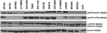
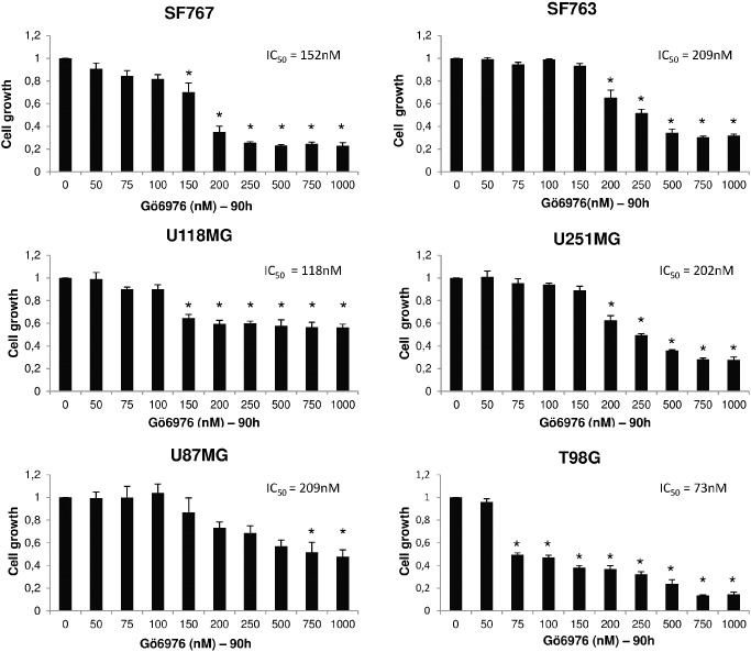
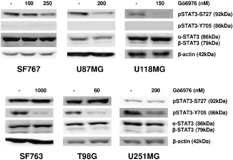
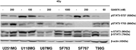
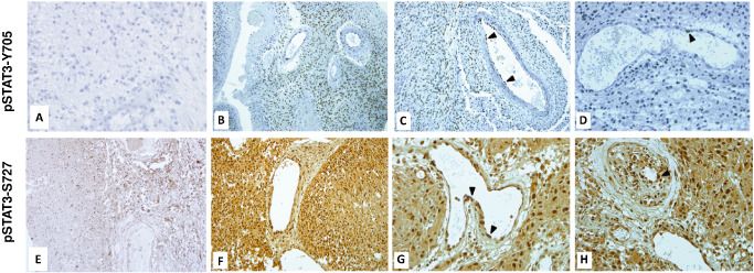
Radiotherapy is an essential component of glioma standard treatment. Glioblastomas (GBM), however, display an important radioresistance leading to tumor recurrence. To improve patient prognosis, there is a need to radiosensitize GBM cells and to circumvent the mechanisms of resistance caused by interactions between tumor cells and their microenvironment. STAT3 has been identified as a therapeutic target in glioma because of its involvement in mechanisms sustaining tumor escape to both standard treatment and immune control. Here, we studied the role of STAT3 activation on tyrosine 705 (Y705) and serine 727 (S727) in glioma radioresistance. This study explored STAT3 phosphorylation on Y705 (pSTAT3-Y705) and S727 (pSTAT3-S727) in glioma cell lines and in clinical samples. Radiosensitizing effect of STAT3 activation down-modulation by Gö6976 was explored. In a panel of 15 human glioma cell lines, we found that the level of pSTAT3-S727 was correlated to intrinsic radioresistance. Moreover, treating GBM cells with Gö6976 resulted in a highly significant radiosensitization associated to a concomitant pSTAT3-S727 down-modulation only in GBM cell lines that exhibited no or weak pSTAT3-Y705. We report the constitutive activation of STAT3-S727 in all GBM clinical samples. Targeting pSTAT3-S727 mainly in pSTAT3-Y705-negative GBM could be a relevant approach to improve radiation therapy.

Keywords: Gö6976; STAT3; glioma; radiotherapy; resistance.


Conflict of interest statement

None.

Figures

Figure 1
Figure 1
STAT3 serine 727 and tyrosine 705 phosphorylation in human malignant glioma cell lines. Cells were harvested during exponential growth phase. Thirty micrograms of total cell lysate were loaded per lane and electrophoresed by SDSPAGE. Transfer membranes were immunoblotted with anti‐STAT3, anti‐pSTAT3Y705 and anti‐pSTAT3S727 specific antibodies. To ensure equal protein loading, β‐actin was used as control. One representative immunoblot is shown. Three independent experiments were achieved for densitometric analyses.
Figure 2
Figure 2
Gö6976 reduces in vitro growth of human glioblastoma. Cells were seeded at 7500 per well of 96‐well plates. Indicated quantity of Gö6976 has been added to growth medium 6 h after seeding. Cells were then grown 90 h before MTT assay. Results represent mean values ± SEM of relative cell growth normalized to untreated cells from three independent experiments done in triplicate; relative IC50 are specified; *significantly different from the control, Dunnett's test.
Figure 3
Figure 3
Gö6976 radiosensitizes pSTAT3Y705‐negative human glioblastoma cells. A–F. Cells were seeded at a density of 5000 per well of 96‐well plates. 6 h later, Gö6976 was added to growth medium. Single 4‐Gy dose irradiation was performed at 24‐h time course incubation with Gö6976. After 90 h of cell growth in Gö6976, cell number was estimated by MTT assay. Results represent mean values ± SEM of relative cell normalized to untreated cells (n = 5, *P < 0.05, Mann–Whitney test). G–I. SF763, SF767 or U118MG cells were incubated 60 h in different concentrations of Gö6976 and PE was determined 10 days after seeding. J–L. For clonogenic survival assay, SF763 and U118MG cells were treated with 1000 nM of Gö6976 and 200 nM for SF767. After 72 h of incubation, cells were irradiated and the surviving fraction was compared with that of control. Data are fitted to linear‐quadratic model. They were represented by their mean ± SEM of values of three independent experiments, each condition performed in triplicate (random‐effects model). Y705‐ : pSTAT3Y705 negative.
Figure 4
Figure 4
Gö6976 down‐modulates pSTAT3S727 only in pSTAT3Y705‐negative human glioblastoma cells. Cells were grown 72 h in indicated concentration of Gö6976 or DMSO (control) before harvest. Thirty micrograms of total proteins were loaded per lane and electrophoresed by SDSPAGE. Transfer membranes were immunoblotted with anti‐STAT3, anti‐pSTAT3Y705 and anti‐pSTAT3S727 specific antibodies. To ensure equal protein loading, β‐actin was used as control.
Figure 5
Figure 5
Subcellular distribution of phosphorylated STAT3 in human glioblastoma cell lines treated by Gö6976. Cells were treated with Gö6976 or DMSO (control) and fixed at 24‐ and 72‐h incubation with Gö6976, respectively, for pSTAT3Y705 and pSTAT3S727 detection. Anti‐pSTAT3Y705 and anti‐pSTAT3‐S727 antibodies were used. DAPI and Cy3‐conjugated secondary goat anti‐rabbit antibody were used. Histograms represent the mean observed intensity of fluorescence in each condition (scale bars are equal to 20 μm, 40×/1.15).
Figure 6
Figure 6
Gö6976 effects after irradiation. Cells were grown 72 h in growth medium with indicated concentration of Gö6976 or DMSO (control) prior to 4‐Gy irradiation. Cells were then harvested 24 h later. Thirty micrograms of total proteins were loaded per lane and electrophoresed by SDSPAGE. Transfer membranes were immunoblotted with anti‐STAT3, anti‐pSTAT3Y705 and anti‐pSTAT3S727 specific antibodies. To ensure equal protein loading, β‐actin was used as control.
Figure 7
Figure 7
STAT3 activation in glioblastoma clinical samples. Immunostaining of pSTAT3 was performed on paraffin‐embedded sections. A. Negative pSTAT3Y705 stained section (×40). B,C. Positive pSTAT3Y705 staining of GBM and vascular endothelial cells (×20). D. Heterogeneous pSTAT3Y705 staining of endothelial cells (×40). E. pSTAT3S727 staining of a transitional zone in a tumor periphery, displaying positively stained GBM cells and unstained non‐neoplastic glial cells (×20). F. pSTAT3S727 staining of GBM cells (×20). G. pSTAT3S727 staining of vascular endothelial cells (×40). H. pSTAT3S727 staining in case of capillary endothelial cell proliferation (×40).

References

    1. Abou‐Ghazal M, Yang DS, Qiao W, Reina‐Ortiz C, Wei J, Kong L‐Y et al (2008) The incidence, correlation with tumor‐infiltrating inflammation, and prognosis of phosphorylated STAT3 expression in human gliomas. Clin Cancer Res 14:8228–8235. - PMC - PubMed
    1. Amberger‐Murphy V (2009) Hypoxia helps glioma to fight therapy. Curr Cancer Drug Targets 9:381–390. - PubMed
    1. Aziz MH, Hafeez BB, Sand JM, Pierce DB, Aziz SW, Dreckschmidt NE, Verma AK (2010) Protein kinase Cε mediates Stat3Ser727 phosphorylation, Stat3‐regulated gene expression and cell invasion in various human cancer cell lines via integration with MAPK cascade (RAF‐1, MEK1/2, and ERK1/2). Oncogene 29:3100–3109. - PMC - PubMed
    1. Barcellos‐Hoff MH, Newcomb EW, Zagzag D, Narayana A (2009) Therapeutic targets in malignant glioblastoma microenvironment. Semin Radiat Oncol 19:163–170. - PMC - PubMed
    1. Bowman T, Garcia R, Turkson J, Jove R (2000) STATs in oncogenesis. Oncogene 19:2474–2488. - PubMed

Publication types

MeSH terms