. 2015 Mar 4:6:6428.
doi: 10.1038/ncomms7428.

PRMT9 is a type II methyltransferase that methylates the splicing factor SAP145

Affiliations

PRMT9 is a type II methyltransferase that methylates the splicing factor SAP145

Yanzhong Yang et al. Nat Commun. .

Abstract

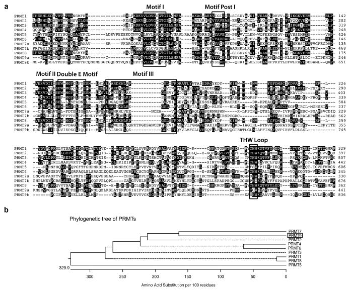
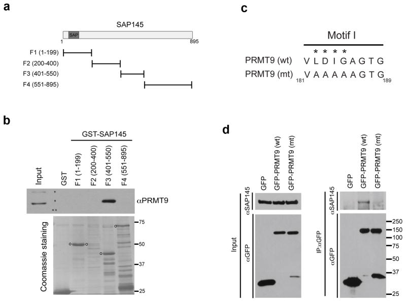
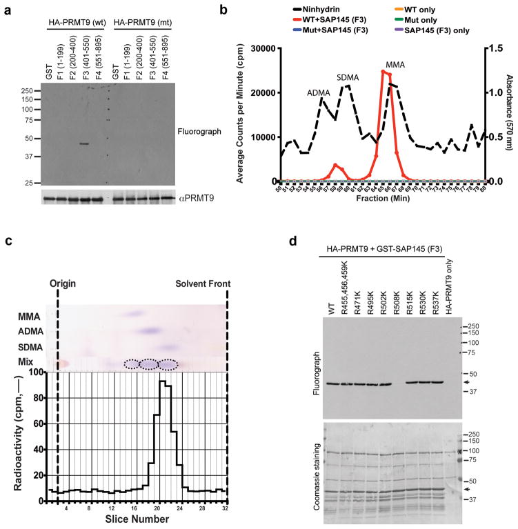
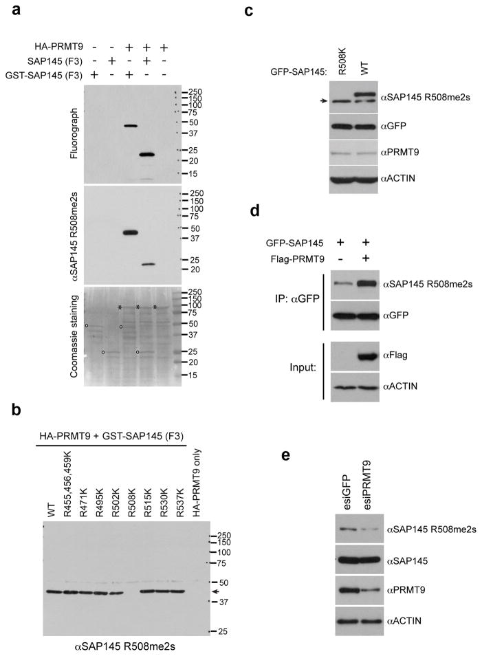
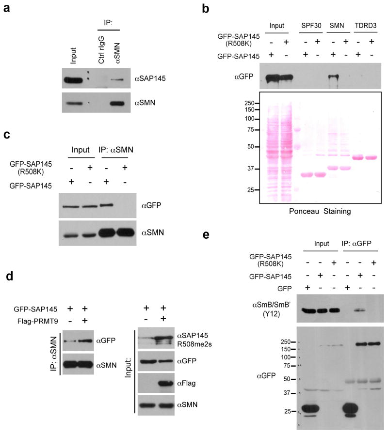
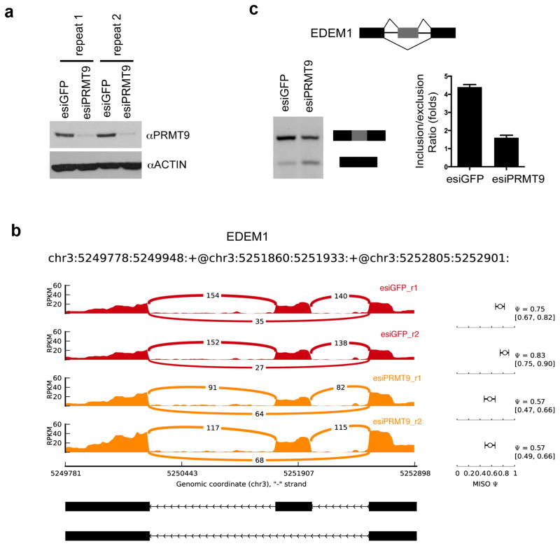
The human genome encodes a family of nine protein arginine methyltransferases (PRMT1-9), whose members can catalyse three distinct types of methylation on arginine residues. Here we identify two spliceosome-associated proteins-SAP145 and SAP49-as PRMT9-binding partners, linking PRMT9 to U2 snRNP maturation. We show that SAP145 is methylated by PRMT9 at arginine 508, which takes the form of monomethylated arginine (MMA) and symmetrically dimethylated arginine (SDMA). PRMT9 thus joins PRMT5 as the only mammalian enzymes capable of depositing the SDMA mark. Methylation of SAP145 on Arg 508 generates a binding site for the Tudor domain of the Survival of Motor Neuron (SMN) protein, and RNA-seq analysis reveals gross splicing changes when PRMT9 levels are attenuated. These results identify PRMT9 as a nonhistone methyltransferase that primes the U2 snRNP for interaction with SMN.


Conflict of interest statement

Competing Financial interests: M.T.B is a cofounder of EpiCypher and a consultant for Cell Signaling Technologies.

Figures

Fig. 1
Fig. 1. Amino acid sequence alignment of human PRMTs
(a) The amino acid sequences from the catalytic domain of PRMTs are compared using ClustalW. The signature sequence motifs are boxed with black squares, including the motifs common to seven beta strand enzymes (Motif I, Motif Post I, Motif II, and Motif III) as well as the Double E Motif and THW Loop motifs that are specific to PRMTs. The number on the right indicates the positions of amino acid of individual PRMT, starting at the initiator methionine. Human PRMT sequences used included PRMT1: NP_001527.3; PRMT2: NP_996845.1; PRMT3: NP_005779.1; PRMT4: NP_954592.1; PRMT5: NP_006100.2; PRMT6: NP_060607.2; PRMT7: NP_061896.1; PRMT8: NP_062828.3; and PRMT9: NP_612373.2. (b) A straight branches phylogenetic tree of all human PRMTs (shown in phenogram) was generated using ClustalW to demonstrate the evolutionary relationships among these enzymes. Protein sequence accession numbers used in the analysis are listed above.
Fig. 2
Fig. 2. PRMT9 interacts with SAP145 and SAP49
(a) TAP-tag purification of PRMT9 protein complex from HeLa cells. HeLa cells were transiently transfected with either empty TAP-tag vector (TAP-Ctrl) or TAP-tag PRMT9 (TAP-PRMT9). A standard TAP procedure was applied. The eluted protein complex was separated by SDS-PAGE and silver-stained. The indicated gel slices were processed for protein identification using mass spectrometry. Actin, heat shock protein 70 (HSP70), and 40S Ribosomal Protein S3 (RS3) are likely non-specific interacting proteins. (b) PRMT9 and SAP145 co-immunoprecipitate. Reciprocal Co-IP was performed in HeLa cells. Total cell lysates were immunoprecipitated with rabbit control IgG, αSAP145 antibody (left), and mouse control IgG, αPRMT9 antibody (right). The eluted protein samples were detected by western blotting with αPRMT9 and αSAP145 antibodies. The input samples were detected with αPRMT9 and αSAP145 antibodies, respectively. (c) PRMT9 and SAP49 co-immunoprecipitate. HeLa cells were transiently transfected with either GFP control vector or GFP-SAP49 plasmids. Total cell lysates were immunoprecipitated with αGFP antibody. The eluted protein samples were detected by western blotting with αPRMT9 and αGFP antibodies. The input samples were detected with αPRMT9. (d) Among all the PRMTs, only PRMT9 interacts with SAP145. HeLa cells were transiently transfected with control GFP vector and GFP-PRMTs (1 through 9). The total cell lysates were immunoprecipitated with αGFP antibody. The eluted protein samples were detected by western blotting with αSAP145, αRPS2 and αGFP antibodies. The input samples were detected with αSAP145 and αRPS2 antibodies.
Fig. 3
Fig. 3. Mapping the interaction regions of PRMT9 and SAP145
(a) A series of GST-fusion truncations of SAP145 were generated. The location of the SAP (SAF-A/B, Acinus and PIAS) domain is indicated. (b) PRMT9 interacts with the amino acids 401–550 of SAP145. GST pull-down experiment was performed by incubating HeLa cell total lysates with purified GST or GST-tag SAP145 fragments described in (A). The pull-down samples were eluted and detected by western blotting using αPRMT9 antibody. The loading of the proteins was visualized by coomassie staining of the membrane. The open circles indicate the individual GST-tag SAP145 fragments. (c) Generation of enzymatic mutant PRMT9. The amino acids (LDIG) located within the conserved Motif I of PRMT9 were mutated to AAAA, causing the loss of methyltransferase activity of enzyme. The numbers below indicate the location of the amino acids on the protein. (d) Wild type, but not the enzymatic mutant, PRMT9 interacts with SAP145. Co-immunoprecipitation was performed in HeLa cells transiently transfected with GFP control vector, GFP-PRMT9 (wt) and GFP-PRMT9 (mt), as described in (c). Total cell lysates were immunoprecipitated with αGFP antibody. The eluted protein samples were detected by western blotting with αSAP145 and αGFP antibodies. The input samples were detected with αSAP145 and αGFP antibodies as well.
Fig. 4
Fig. 4. PRMT9 catalyzes symmetrical dimethylation of SAP145 at Arginine 508
(a) PRMT9 methylates SAP145 fragment F3 (a.a. 401–550). The in vitro methylation was performed by incubating either wild type or enzymatic mutant recombinant HA-PRMT9 (purified from Sf21 cells) with GST or GST-tag SAP145 fragments (F1–F4, as described in Fig. 3a and b). The loading of PRMT9 was detected by western blotting using αHA antibody. (b) PRMT9 symmetrically dimethylates SAP145 as detected by amino acid analysis. Amino acid analysis of in vitro methylation products from wild type and enzymatic mutant GFP-PRMT9 as enzymes and GST-SAP145 (401–550) fragment as substrate. Black dashed line indicates elution of nonradiolabeled standards. The radioactive peaks elute 1–2 min before the nonradiolabeled standards due to a tritium isotope effect. (c) PRMT9 symmetrically dimethylates SAP145 as detected by thin layer chromatography (TLC). SDMA fractions from cation-exchange chromatography of the in vitro 3H-methylation reaction were separated using thin layer chromatography (TLC). Fractions were spotted on a cellulose plate along with the addition of 5 nmol MMA, 15 nmol ADMA, and 5 nmol SDMA internal standards. Individual standards were also spotted in adjacent lanes to determine the migration distance of each methylated arginine derivative. The origin is indicated by fraction 2. The solvent front was run near the end of the plate to fraction 32. The plate was air-dried and each lane was subsequently sliced in 5 mm fractions and counted for three 30-min counting cycles. The radioactive peaks elute 1–2 min before the nonradiolabeled standards due to a tritium isotope effect. This experiment was repeated 3 times and similar migration patterns were observed. (d) PRMT9 methylates SAP145 at R508 in vitro. The in vitro methylation assay was performed by incubating recombinant HA-PRMT9 with a series of Arg to Lys (R to K) mutants of SAP145 fragment F3 (see Fig. 3a and b for description) for 1 h at 30° C. After exposure at −80 °C for 3 days, the membrane was stained with Coomassie blue to check the protein loading. Arrows indicate the positions of the substrates and stars indicate the positions of the recombinant HA-PRMT9.
Fig. 5
Fig. 5. PRMT9 symmetrically methylates SAP145 at Arg 508 in vivo
(a) SAP145 R508 methylation specific antibody detects in vitro methylated SAP145. The in vitro methylation assay was performed using recombinant HA-PRMT9 as enzyme, either GST-tag SAP145 fragment F3, or SAP145 fragment F3 without GST-tag (cleaved by PreScissionTM Protease) as substrates. The samples were run on a SDS-PAGE gel followed by exposure to x-ray film for 3 days. After exposure, the membrane was stripped and detected using SAP145 R508 methylation specific antibody (αSAP145 R508me2s) by western blotting. The membrane was then stained with Coomassie blue to check the protein loading. Open circles indicate the positions of the substrates and stars indicate the position of the recombinant HA-PRMT9. (b) Arg to Lys mutation at R508 site of SAP145 abolishes the recognition of αSAP145 R508me2s antibody. The same membrane, described in Fig. 4 (d), was stripped and detected using αSAP145 R508me2s antibody by western blotting. The arrow indicates the position of specific signal detected by the antibody. (c) Arg 508 of SAP145 is symmetrically dimethylated in the cells. HeLa cells were transiently transfected with either GFP-SAP145 or GFP-SAP145 (R508K). 30 μg of total cell lysates were subjected to western blotting detection with αSAP145 R508me2s, αGFP, αPRMT9 and αActin antibodies. The arrow indicates the endogenous SAP145 methylation signals. (d) Overexpression of PRMT9 increases SAP145 R508 symmetrical dimethylation. HeLa cells were transfected with either GFP-SAP145 alone, or GFP-SAP145 and Flag-PRMT9. The total cell lysates were immunoprecipitated with αGFP antibody followed by western blotting with αSAP145 R508me2s and αGFP antibodies. The input samples were detected with αFlag and αActin antibodies. (e) Knockdown of PRMT9 decreases SAP145 R508 symmetric dimethylation. HeLa cells were transiently transfected with either control esiGFP (esiRNA targeting GFP) or esiPRMT9 (esiRNA targeting PRMT9). 30 μg of total cell lysates were subjected to western blotting and detected with αSAP145 R508me2s, αSAP145, αPRMT9 and αActin antibodies.
Fig. 6
Fig. 6. Methylation of Arginine 508 is required for SAP145-SMN interaction
(a) SMN interacts with SAP145 in vivo. Endogenous Co-IP was performed by immunoprecipitating HeLa cell nuclear extracts using αSMN antibody, followed by western blotting detection using αSAP145 antibody. (b) Tudor domain of SMN interacts with wild type but not R508K mutant form of SAP145. HeLa cell were transfected with either GFP-SAP145 or GFP-SAP145 (R508K). GST pull-down experiment was performed by incubating transfected cell lysates with recombinant GST-Tudor domains of SPF30, SMN and TDRD3. Eluted samples were detected by western blotting with αGFP antibody. Ponceau S staining displays the loading of total cell lysates and recombinant proteins (c) SMN interacts with the wild type but not the R508K mutant form of SAP145. HeLa cells were transfected with either GFP-SAP145 or GFP-SAP145 (R508K). The nuclear extracts were immunoprecipitated with αSMN antibody. The eluted samples and input samples were detected with the indicated antibodies. (d) Overexpression of PRMT9 enhances the interaction of SMN with GFP-SAP145. HeLa cells were transfected with the indicated plasmids. The nuclear extracts were immunoprecipitated with αSMN antibody. The eluted samples and input samples were detected with the indicated antibodies. (e) An intact SAP145 R508 methylation site is required for U2 snRNP maturation. HeLa cells were transfected with either GFP-SAP145 or GFP-SAP145 (R508K). Cell lysates were immunoprecipitated with αGFP antibody. The eluted samples and input samples were detected with αSmB (Y12) and with αGFP antibodies.
Fig. 7
Fig. 7. PRMT9 regulates alternative splicing
(a) Two sets of biological replicated samples for RNA-seq. HeLa cells were transiently transfected with esiGFP control or esiPRMT9. The total RNA was extracted 72 hours after transfection. Western blotting confirms the knockdown of PRMT9 protein using αPRMT9 and αActin antibodies. (b) RNA-seq read coverage across EDEM1 exon 8 to exon 10 from esiGFP control and esiPRMT9 knockdown HeLa cells. MISO (Ψ) values and 95% confidence intervals were shown at right. (c) Validation of exon exclusion of EDEM1 exon 9 in response to PRMT9 knockdown. RT-PCR experiments were performed using RNA extracted from esiGFP control and esiPRMT9 transfected HeLa cells with primers located on exon 8 and exon 10 (left panel). The PCR products were quantified by densitometric analysis. Inclusion vs exclusion ratio was calculated. Error bars represent standard deviation calculated from three independent experiments (right panel).

Similar articles

Cited by

References

    1. Dhar S, et al. Loss of the major Type I arginine methyltransferase PRMT1 causes substrate scavenging by other PRMTs. Sci Rep. 2013;3:1311. - PMC - PubMed
    1. Gary JD, Clarke S. RNA and protein interactions modulated by protein arginine methylation. Prog Nucleic Acid Res Mol Biol. 1998;61:65–131. - PubMed
    1. Liu Q, Dreyfuss G. In vivo and in vitro arginine methylation of RNA-binding proteins. Mol Cell Biol. 1995;15:2800–2808. - PMC - PubMed
    1. Boffa LC, Karn J, Vidali G, Allfrey VG. Distribution of NG, NG,-dimethylarginine in nuclear protein fractions. Biochem Biophys Res Commun. 1977;74:969–976. - PubMed
    1. Gayatri S, Bedford MT. Readers of histone methylarginine marks. Biochimica et biophysica acta. 2014 doi: 10.1016/j.bbagrm.2014.02.015. - DOI - PMC - PubMed

Publication types

MeSH terms