Review
. 2015 Apr 15;209(1-2):1-42.
doi: 10.1016/j.vetpar.2015.01.026. Epub 2015 Feb 7.

An update on Sarcocystis neurona infections in animals and equine protozoal myeloencephalitis (EPM)

Affiliations
Review

An update on Sarcocystis neurona infections in animals and equine protozoal myeloencephalitis (EPM)

J P Dubey et al. Vet Parasitol. .

Abstract

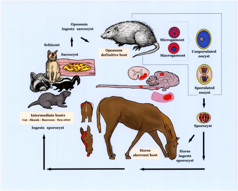
Equine protozoal myeloencephalitis (EPM) is a serious disease of horses, and its management continues to be a challenge for veterinarians. The protozoan Sarcocystis neurona is most commonly associated with EPM. S. neurona has emerged as a common cause of mortality in marine mammals, especially sea otters (Enhydra lutris). EPM-like illness has also been recorded in several other mammals, including domestic dogs and cats. This paper updates S. neurona and EPM information from the last 15 years on the advances regarding life cycle, molecular biology, epidemiology, clinical signs, diagnosis, treatment and control.

Keywords: Epidemiology; Equine protozoal myeloencephalitis; Life cycle; Marine mammals; Prevention; Sarcocystis neurona.


Figures

Figure 1
Figure 1
Life cycle of S. neurona. Opossums are the definitive host and other animals are aberrant/intermediate hosts. S. neurona parasitizes and causes lesions (in red) in the brain and spinal cord of horses. Affected horses can have neurological signs, including abnormal gait, dysphagia, and muscle atrophy depicted.
Figure 2
Figure 2
Fluorescence images of S. neurona. (A) Images of gliding S. neurona merozoites stained with a monoclonal antibody (2A7-18) to the surface protein of Sn-SAG1. The formation of the trails is similar to those reported for Toxoplasma gondii. The trails are readily visualized by staining with antibodies to the major surface antigens. Gliding occurs on a variety of substrates, including coated chamber slides (50% PBS and fetal bovine serum). (B) Transgenic clone of Sarcocystis neurona expressing yellow fluorescent protein. Differential interference contrast image with epifluorescence image overlay showing a bovine turbinate cell monolayer containing a late-stage schizont and a mature schizont of a clone of S. neurona that stably expresses YFP. Host cell and parasite nuclei were stained with DAPI (blue). Bar= 10μm.
Figure 3
Figure 3
S. neurona schizonts and merozoites in tissue smears. Giemsa stain. (A) Smear from a pustular dermal ulcer from a Rottweiler dog reported by Dubey et al. (1991b). Note several intensely stained schizonts (arrows) among leukocytes and RBC. (B) Infected dermal cell with immature schizonts (sc) and free elongated merozoites compared with size or RBC. Note host cell nucleus (hcn). (C) Smear of brain of a red panda. Note two different sized merozoites (a), a merozoite-shaped schizont with lobed nucleus (b), and two schizonts with large nucleus (c), and an intensely stained schizont (d). (Smear courtesy of Timothy Walsh, unpublished).
Figure 4
Figure 4
S. neurona -associated lesions in the spinal cord of a dog reported by Gerhold et al. (2014). (A) Nodular growth (arrow) between spinal nerve roots. Unstained. Bar =1cm. (B) Cut section of the spinal cord in Fig. 11A. The protruding lesion is extended in to the central canal. Unstained. C to F histological sections. C and D, hematoxylin and eosin stain, E and F immunohistochemical staining with S. neurona antibody. Bar in C, D, and F=5 μm, and in E=20 μm. (C) Note different developing schizogonic stages in one field-merozoite (a), an immature schizont with multilobed nucleus (b), schizonts with developing merozoites (c,d). (D) At least 7 schizonts within a phagocytic cell. Arrow points to a mature schizont with residual body. Arrowheads point to immature schizonts. (E) Numerous free merozoites and schizonts. The organisms appear larger in size after immunostaining. (F) A phagocytic host cell (arrow) similar to that in Fig. 11D showing several schizonts (specimens courtesy of Shelley J Newman, and Amanda Crews).
Figure 5
Figure 5
TEM of an infected neural cell in the brain of a raccoon naturally infected with S. neurona. Note asynchronous schizogony with six developing schizonts, in presumed order of development (a-e), and nine merozoites (f). Arrow points to a longitudinally merozoite with a conoid at one end and a subterminal nucleus. The host cell is degerated but parasite structures are fairly well preserved (From Dubey et al., 1991c).
Figure 6
Figure 6
TEM of a bovine turbinate cell culture infected with S. neurona. Mature schizont with non conoidal end of merozoites still attached to a residual body (rb). Note one longitudinally cut merozoite (arrow) that has separated from the schizont, and the host cell nucleus (hcn). (From Dubey, 2004).
Figure 7
Figure 7
S. neurona merozoites free in cytoplasm of an unmyelinated axon in the cerebellum of an experimentally infected KO mouse. (From Fritz and Dubey, 2002).
Figure 8
Figure 8
S. neurona sarcocysts in histological sections of skeletal muscle. Arrowheads point to striated cyst wall. Arrows point to thickening of the villar tips. (A) Cat, 144 DPI. From Dubey et al., 2002).Toluidine blue stain. (B) Raccoon, 77 DPI. Hematoxylin and eosin. (C) Toluidine blue stain. (B and C from Stanek et al., 2002).
Figure 9
Figure 9
Comparison of the cyst walls of S. neurona (A) and S. fayeri (B) sarcocysts by TEM. The cyst walls, including the ground substance layer (gs) of S. fayeri are thick, the microtubules (mt) are more electrondense and extend up to the pellicle of the zoites whereas the cyst walls of S. neurona are comparatively thin, the microtubules are few, and never extend deep in the gs. (From Stanek et al., 2002, and Saville et al., 2004b).
Figure 10
Figure 10
Comparison of a merozoite and a bradyzoite of S. neurona. (A) Merozoite in the brain of a naturally infected horse (From Dubey et al., 1998). (B) Bradyzoite in sarcocyst in an experimentally infected cat (From Dubey et al., 2001d). Note the location of nucleus (nu), central in merozoite, and terminal in bradyzoite, and the absence of rhoptries in merozoite. Also note conoid (co), micronemes (mn) and dense granules (dg). Dense granules are often mistaken for rhoptries unless their elongated portions are visible.
Figure 11
Figure 11
Two horses with clinical EPM. Top, ataxic and bottom a mare with urinary inconsitance.
Figure 12
Figure 12
Surface (top) and cut (bottom) views of cerebrum of a 20 year old Paint horse with histologically and PCR confirmed EPM. The horse had a six day history of muscle fasciculations, bruxism, difficulty eating and drinking, and circling to the left with head pressing. Note hemorrhagic and yellow discolored areas indicative of necrosis. Bar = 5 mm. (Courtesy of Uneeda Bryant).

References

    1. Arias M, Yeargan M, Francisco I, Dangoudoubiyam S, Becerra P, Francisco R, Sánchez-Andrade R, Paz-Silva A, Howe DK. Exposure to Sarcocystis spp. in horses from Spain determined by Western blot analysis using Sarcocystis neurona merozoites as heterologous antigen. Vet Parasitol. 2012;185:301–304. - PubMed
    1. Asai T, Takeuchi T, Diffenderfer J, Sibley LD. Identification of small-molecule inhibitors of nucleoside triphosphate hydrolase in Toxoplasma gondii. Antimicrob Agents Chemother. 2002;46:2393–2399. - PMC - PubMed
    1. Asmundsson IM, Dubey JP, Rosenthal BM. A genetically diverse but distinct North American population of Sarcocystis neurona includes an overrepresented clone described by 12 microsatellite alleles. Infect Genet Evol. 2006;6:352–360. - PubMed
    1. Asmundsson IM, Rosenthal BM. Isolation and characterization of microsatellite markers from Sarcocystis neurona, a causative agent of equine protozoal myeloencephalitis. Molecular Ecology Notes. 2006;6:8–10.
    1. Barr SC, Warner K. Characterization of a serine protease activity in Sarcocystis neurona merozoites. J Parasitol. 2003;89:385–388. - PubMed

Publication types

MeSH terms

Substances

LinkOut - more resources