

The composition of cellular infiltrates in anti-HMG-CoA reductase-associated myopathy
- PMID: 25737145
- PMCID: PMC4510021
- DOI: 10.1002/mus.24642
The composition of cellular infiltrates in anti-HMG-CoA reductase-associated myopathy
Abstract
Introduction: To characterize cellular infiltrates in muscle biopsies from patients with anti-3-hydroxy-3-methyl-gulatryl-CoA reductase (HMGCR)-associated myopathy.
Methods: Biopsies from 18 anti-HMGCR myopathy and 7 control dermatomyositis patients were analyzed.
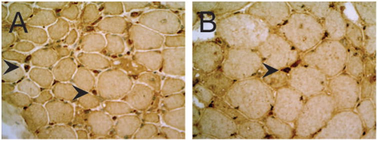
Results: CD4+ and CD8+ T-cells were scattered within the endomysium in 50% of anti-HMGCR biopsies. All anti-HMGCR biopsies included increased endomysial and/or perivascular CD163+ M2 macrophages; CD11c+ M1 macrophages were present in 18.8%. CD123+ plasmacytoid dendritic (PD) cells were observed within the endomysium and perivascular spaces in 62.5% of anti-HMGCR biopsies. Membrane attack complex was deposited on endothelial cells in 50% and on the sarcolemma of nonnecrotic muscle fibers in 85.7% of anti-HMGCR cases. Major histocompatibility complex class I antigen was up-regulated in 87.5% of the anti-HMGCR cases.
Conclusions: In addition to necrosis, scattered CD4+, CD8+, and PD cells are characteristic of anti-HMGCR myopathy. Predominant M2 polarization suggests infiltrating macrophages are more likely to be involved with tissue repair than destruction.
Keywords: anti-HMGCR myopathy; autoimmune myopathy; immune-mediated necrotizing myopathy; muscle histology; myositis.
© 2015 Wiley Periodicals, Inc.
Conflict of interest statement
Figures


References
-
- Mammen AL. Dermatomyositis and polymyositis: Clinical presentation, autoantibodies, and pathogenesis. Ann N Y Acad Sci. 2010;1184:134–153. - PubMed
-
- Hoogendijk JE, Amato AA, Lecky BR, Choy EH, Lundberg IE, Rose MR, et al. 119th ENMC international workshop: trial design in adult idiopathic inflammatory myopathies, with the exception of inclusion body myositis, 10-12 October 2003, Naarden, The Netherlands. Neuromuscul Disord. 2004;14:337–345. - PubMed
-
- Greenberg SA, Pinkus JL, Pinkus GS, Burleson T, Sanoudou D, Tawil R, et al. Interferon-alpha/beta-mediated innate immune mechanisms in dermatomyositis. Ann Neurol. 2005;57:664–678. - PubMed
Publication types
MeSH terms
Substances
Grants and funding
LinkOut - more resources
Full Text Sources
Other Literature Sources
Medical
Research Materials
