Review
. 2015 Mar 1;29(5):467-82.
doi: 10.1101/gad.256396.114.

The yin and yang of kidney development and Wilms' tumors

Affiliations
Review

The yin and yang of kidney development and Wilms' tumors

Peter Hohenstein et al. Genes Dev. .

Abstract

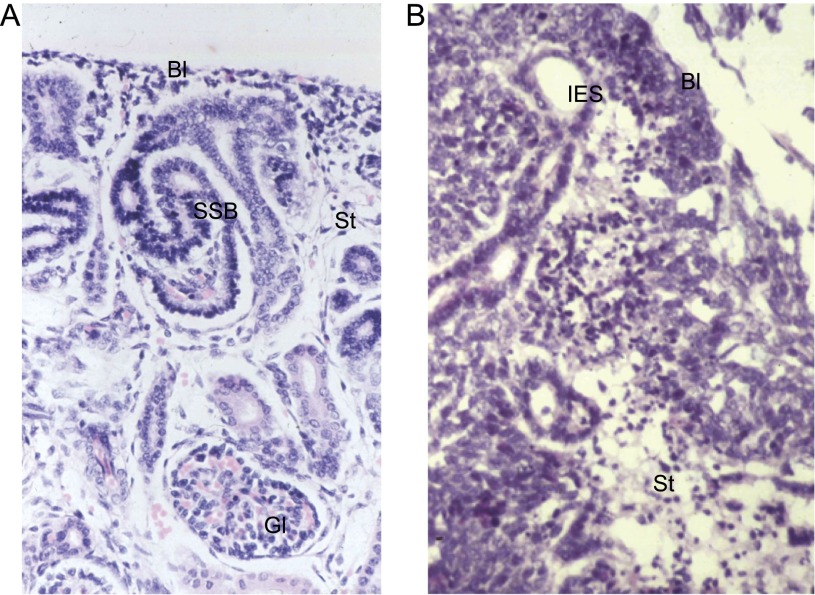
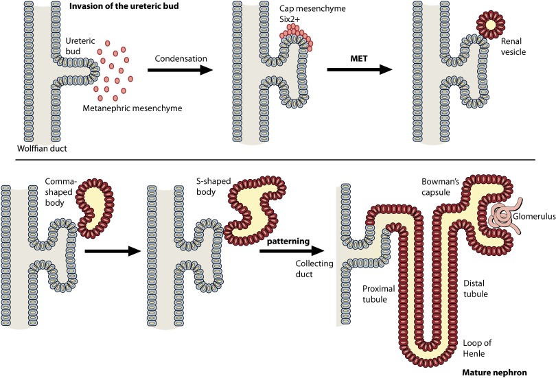
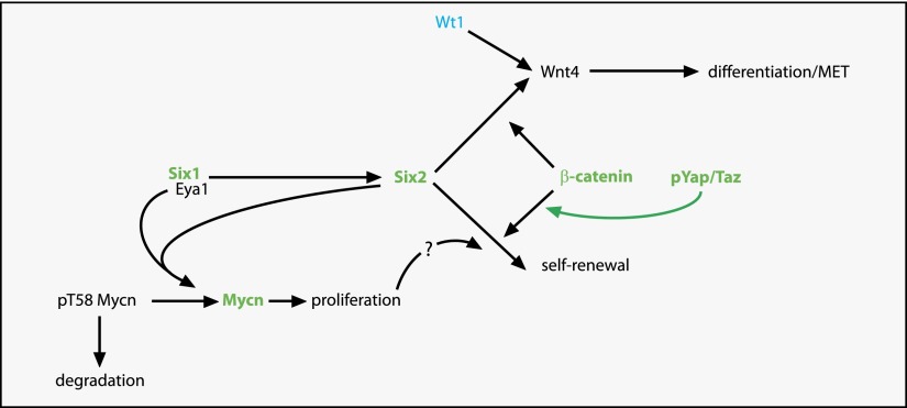
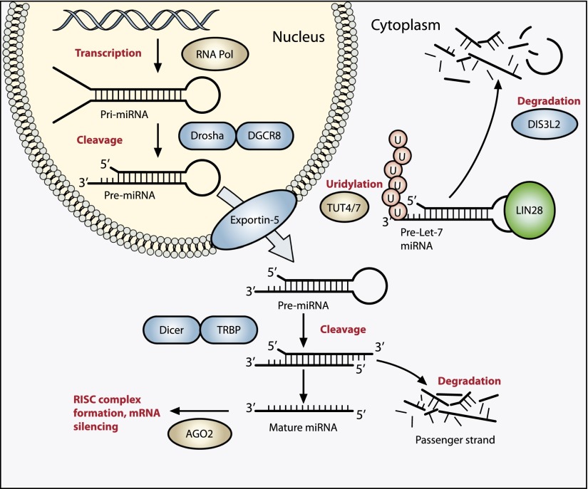
Wilms' tumor, or nephroblastoma, is the most common pediatric renal cancer. The tumors morphologically resemble embryonic kidneys with a disrupted architecture and are associated with undifferentiated metanephric precursors. Here, we discuss genetic and epigenetic findings in Wilms' tumor in the context of renal development. Many of the genes implicated in Wilms' tumorigenesis are involved in the control of nephron progenitors or the microRNA (miRNA) processing pathway. Whereas the first group of genes has been extensively studied in normal development, the second finding suggests important roles for miRNAs in general-and specific miRNAs in particular-in normal kidney development that still await further analysis. The recent identification of Wilms' tumor cancer stem cells could provide a framework to integrate these pathways and translate them into new or improved therapeutic interventions.

Keywords: Wilms’ tumor; Wt1; kidney development; miRNA; nephron progenitor cells; β-catenin.


Figures

Figure 1.
Figure 1.
Wilms’ tumors resemble fetal kidneys. (A) Human fetal kidney (18-wk gestation) showing blastema (metanephric mesenchyme), epithelial differentiation (S-shaped body), stroma, and a mature glomerulus. (B) A triphasic Wilms’ tumor showing all three cell types: blastema, epithelia, and stroma. (Bl) Blastema; (SSB) S-shaped body; (St) stroma; (Gl) glomerulus; (IES) immature epithelial structure.
Figure 2.
Figure 2.
Schematic representation of kidney development. The ureteric bud (gray) extends from the Wolffian duct, and, upon contact with the metanephric mesenchyme (red), reciprocal signaling induces both bud bifurcation and condensation of the mesenchyme to generate the cap mesenchyme. The blastemal mesenchyme then undergo a MET to generate the renal vesicle, which continues to form the comma-shaped and S-shaped body before the distal end fuses with the ureteric bud (which forms the collecting duct), and the proximal end joins to form the glomerulus, generating the mature nephron (dark orange).
Figure 3.
Figure 3.
Control of nephron progenitor cells is disturbed in many Wilms’ tumors. Model for the control of the fate of nephron progenitor cells. Eya1 lies genetically upstream of Six2. Six2 labels the nephron progenitor cells, which can either maintain a progenitor state and self-renew or differentiate via the Wnt4-mediated MET. Wnt4 expression is under the direct control of Wt1. β-Catenin is involved in both progenitor cell fates through activation of different transcriptional programs. Active nuclear phosphorylated Yap/Taz shifts the progenitor balance toward the self-renewal fate. Eya1 and Six2 interact directly with Mycn, leading to dephosphorylation of Mycn pT58, stabilization of the protein, increased proliferation, and potentially a shift of the nephron progenitor toward self-renewal. Genes activated in Wilms’ tumors are depicted in green, and inactivated genes are in blue. Deregulation of Yap/Taz in Wilms’ tumors results in phosphorylated Yap not being retained in the cytoplasm as it should, but it translocates to the nucleus and thus shifts the progenitor cell balance toward self-renewal. This model is likely a simplification, as it presumes that all Wilms’ tumors, regardless of causative mutation, are caused by the same mechanism.
Figure 4.
Figure 4.
The miRNA processing pathway is commonly mutated in Wilms’ tumor. Expression of mature miRNA is initiated by RNA polymerase-mediated transcription of DNA-encoded sequences into pri-miRNA, which form a long double-stranded hairpin. This structure is then cleaved by a complex of Drosha and DGCR8 (termed Pasha) into a smaller pre-miRNA hairpin, which is exported from the nucleus and then cleaved by Dicer (an RNase) and TRBP (with specificity for dsRNA) to remove the hairpin loop and leave two single-stranded miRNAs. The functional strand binds to Argonaute (Ago2) proteins into the RNA-induced silencing complex (RISC), where it guides the complex to its target mRNA, while the nonfunctional strand is degraded. Targeting of mRNAs by this method results in mRNA silencing by mRNA cleavage, translational repression, or deadenylation. Let-7 miRNAs are a family of miRNAs highly expressed in ESCs with tumor suppressor properties. In cases in which LIN28 is overexpressed, LIN28 binds to pre-Let-7 miRNA, preventing DICER from binding and resulting in LIN28-activated polyuridylation by TUT4 or TUT7, causing reciprocal DIS3L2-mediated degradation of Let-7 pre-miRNAs. Genes involved in miRNA processing that have been associated with Wilms’ tumor are highlighted in blue (inactivating) and green (activating) and include DROSHA, DGCR8, XPO5 (encoding exportin-5), DICER1, TARBP2, DIS3L2, and LIN28.

References

    1. Aiden AP, Rivera MN, Rheinbay E, Ku M, Coffman EJ, Truong TT, Vargas SO, Lander ES, Haber DA, Bernstein BE. 2010. Wilms tumor chromatin profiles highlight stem cell properties and a renal developmental network. Cell Stem Cell 6: 591–602. - PMC - PubMed
    1. Anglesio MS, Evdokimova V, Melnyk N, Zhang L, Fernandez CV, Grundy PE, Leach S, Marra MA, Brooks-Wilson AR, Penninger J, et al. . 2004. Differential expression of a novel ankyrin containing E3 ubiquitin-protein ligase, Hace1, in sporadic Wilms’ tumor versus normal kidney. Hum Mol Genet 13: 2061–2074. - PubMed
    1. Astuti D, Morris MR, Cooper WN, Staals RH, Wake NC, Fews GA, Gill H, Gentle D, Shuib S, Ricketts CJ, et al. . 2012. Germline mutations in DIS3L2 cause the Perlman syndrome of overgrowth and Wilms tumor susceptibility. Nat Genet 44: 277–284. - PubMed
    1. Bahubeshi A, Bal N, Rio Frio T, Hamel N, Pouchet C, Yilmaz A, Bouron-Dal Soglio D, Williams GM, Tischkowitz M, Priest JR, et al. . 2010. Germline DICER1 mutations and familial cystic nephroma. J Med Genet 47: 863–866. - PubMed
    1. Bardeesy N, Falkoff D, Petruzzi M-J, Nowak N, Zabel B, Adam M, Aguiar MC, Grundy P, Shows T, Pelletier J. 1994. Anaplastic Wilms’ tumour, a subtype displaying poor prognosis, harbours p53 gene mutations. Nat Genet 7: 91–97. - PubMed

Publication types

LinkOut - more resources