. 2015 Feb 28;6(6):4239-52.
doi: 10.18632/oncotarget.2978.

WISP-1 a novel angiogenic regulator of the CCN family promotes oral squamous cell carcinoma angiogenesis through VEGF-A expression

Affiliations

WISP-1 a novel angiogenic regulator of the CCN family promotes oral squamous cell carcinoma angiogenesis through VEGF-A expression

Jing-Yuan Chuang et al. Oncotarget. .

Abstract

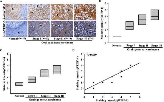
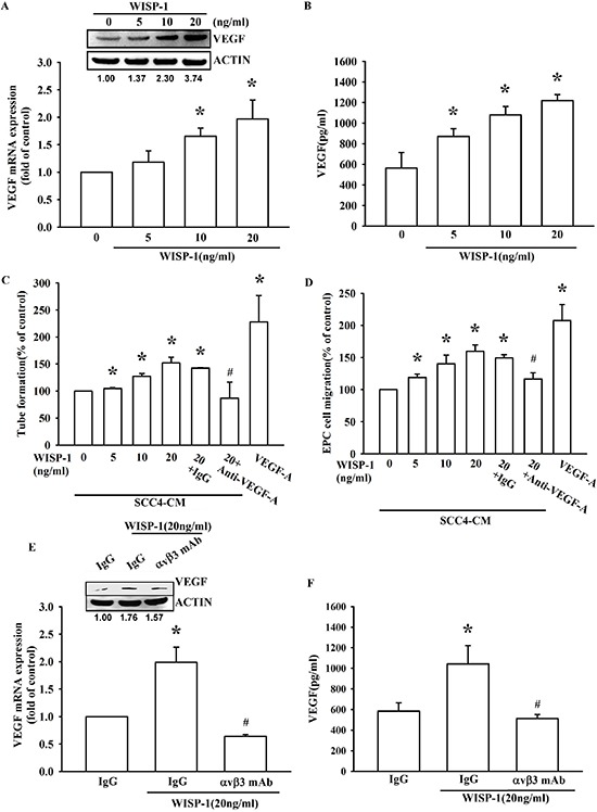
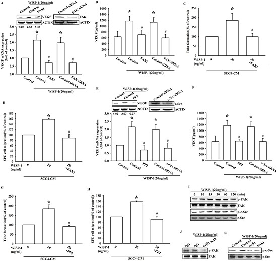
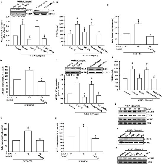
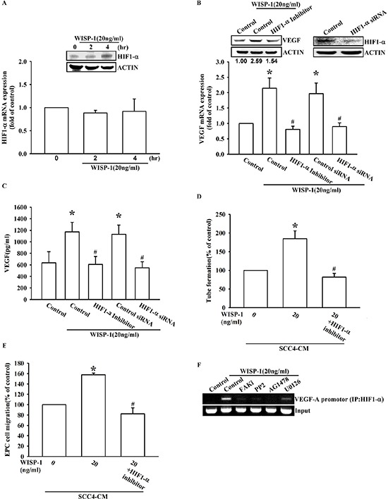
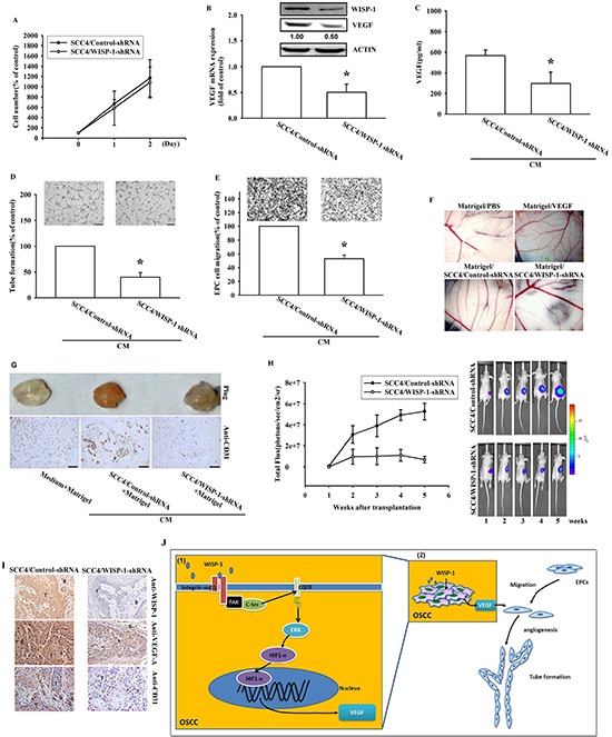
Oral squamous cell carcinoma (OSCC), which accounts for nearly 90% of head and neck cancers, is characterized by poor prognosis and a low survival rate. VEGF-A is the most established angiogenic factor involved in the angiogenic-regulated tumor progression. WISP-1/CCN4 is an extracellular matrix-related protein that belongs to the Cyr61, CTGF, Nov (CCN) family and regulates many biological functions, such as angiogenesis. Previous studies indicated the role of WISP-1 in tumor progression. However, the angiogenic property of WISP-1 in the cancer microenvironment has never been discussed. Here, we provide novel insights regarding the role of WISP-1 in the angiogenesis through promoting VEGF-A expression. In this study, the correlation of WISP-1 and VEGF-A was confirmed by IHC staining of specimens from patients with OSCC. In vitro results indicated that WISP-1 induced VEGF-A expression via the integrin αvβ3/FAK/c-Src pathway, which transactivates the EGFR/ERK/HIF1-α signaling pathway in OSCC. This pathway in turn induces the recruitment of endothelial progenitor cells and triggers the neovascularization in the tumor microenvironment. Our in vivo data revealed that tumor-secreted WISP-1 promoted the angiogenesis through VRGF expression and increased angiogenesis-related tumor growth. Our study offers new information that highlights WISP-1 as a potential novel therapeutic target for OSCC.


Conflict of interest statement

Conflict of Interest

The authors declare that they have no competing interests.

Figures

Figure 1
Figure 1. Clinical significance of WISP-1 and VEGF-A in specimens from patients with OSCC
Tumor specimens were immunostained (IHC) with anti-WISP-1 and anti-VEGF-A antibodies. The staining intensity was scored 1–5. (A) IHC photographs. (E = epithelial, T = tumor, S = stroma). (B–D) Quantitative results and correlation between WISP-1, VEGF-A, and OSCC clinical grade.
Figure 2
Figure 2. WISP-1 regulates the angiogenesis by raising VEGF-A expression in OSCC cells
(A–B) SCC4 cells were incubated with WISP-1 (0–20 ng/mL) for 24 h, VEGF-A expression was measured by qPCR, ELISA, and western blot. (C–D) SCC4 cells were incubated with WISP-1 (0–20 ng/mL) for 24 h, and the CM was collected. EPCs were pre-treated for 30 min with IgG control antibody or VEGF-A antibody (1 μg/mL) and incubated with CM for 6 h and cell capillary-like structure formation in EPCs was examined by tube formation assay (C) EPCs were incubated with CM for 24 h, and cell migration was examined using the transwell assay (D) (E–F) SCC4 cells were incubated with the integrin αvβ3 antibody for 30 min, followed by stimulation with WISP-1 (20 ng/mL) for 24 h. VEGF-A expression was examined by western blot, qPCR, and ELISA. Data are expressed as mean ± SEM *P < 0.05 compared to control; #P < 0.05 compared to the WISP-1 treated group.
Figure 3
Figure 3. FAK/Src signaling pathway is involved in WISP-1-promoted VEGF-A expression and contributing to angiogenesis
(A–B) SCC4 cells were pre-treated with a FAK inhibitor (FAKi; 1 μM) for 30 min or transfected with FAK siRNAs for 24 h, followed by WISP-1 (20 ng/mL) stimulation for 24 h. VEGF-A expression was examined by western blot, qPCR, and ELISA. (C–D) SCC4 cells were pre-treated with a FAK inhibitor (FAKi; 1 μM), followed by WISP-1 (20 ng/mL) stimulation for 24 h. CM was collected. EPCs were incubated with CM for 6 h and capillary-like structure formation in EPCs was examined by tube formation assay (C) EPCs were incubated with CM for 24 h and cell migration was examined by transwell assay (D) (E–H) SCC4 cells were treated with a Src inhibitor (pp2; 1 μM) for 30 min or transfected with Src siRNAs for 24 h, followed by WISP-1 (20 ng/mL) stimulation for 24 h. Assay procedure was performed as in (A–D) (I) SCC4 cells were incubated with WISP-1 (20 ng/mL) for the indicated times, and FAK and c-Src phosphorylation was determined by western blot. (J–K) SCC4 cells were incubated with an integrin αvβ3 antibody or FAKi for 30 min, followed by stimulation with WISP-1 (20 ng/mL) for 60 min, and FAK (J) and c-Src (K) phosphorylation was determined by western blot. Data are expressed as the mean ± SEM *P < 0.05 compared with control; #P < 0.05 compared with the WISP-1-treated group.
Figure 4
Figure 4. EGFR transactivation is involved in WISP-1-induced VEGF-A expression and contributing to angiogenesis
(A–D) SCC4 cells were pre-treated with an EGFR inhibitor (AG1478; 1 μM) for 30 min or transfected with EGFR siRNAs for 24 h, followed by WISP-1 (20 ng/mL) stimulation for 24 h. The assay procedures were performed as described in Figure 3A–3D. (E–H) SCC4 cells were treated by an ERK inhibitor (U0126; 1 μM) for 30 min or transfected with ERK siRNAs for 24 h, followed by WISP-1 (20 ng/mL) stimulation for 24 h. The assay procedures were performed as described in Figure 3A–3D. (I) SCC4 cells were incubated with WISP-1 (20 ng/mL) for the indicated times and EGFR and ERK phosphorylation was determined by western blot. (J–K) SCC4 cells were incubated with the integrin αvβ3 antibody, FAKi, PP2, or AG1478 for 30 min, followed by stimulation with WISP-1 (20 ng/mL) for 60 min, and EGFR (J) and ERK (K) phosphorylation was determined by western blot. Data are expressed as the mean ± SEM *P < 0.05 compared with control; #P < 0.05 compared with the WISP-1-treated group.
Figure 5
Figure 5. WISP-1 promotes VEGF-A expression in OSCC and contributing to angiogenesis through the HIF1-α signaling pathway
(A) SCC4 cells were stimulated by WISP-1 (20 ng/mL) for the indicated times (0, 2, and 4 h). HIF1-α expression level was measured by western blot and qPCR. (B–E) SCC4 cells were pre-treated with HIF1-α inhibitor (1 μM) for 30 min or transfected with HIF1-α siRNAs for 24 h, followed by WISP-1 (20 ng/mL) stimulation for 24 h. The assay procedures were performed as described in Figure 3A–3D. (F) SCC4 cells were incubated with FAKi, PP2, AG1478, or U0126 for 30 min, followed by stimulation with WISP-1 (20 ng/mL) for 60 min. Chromatin immunoprecipitation (ChIP) assays were performed using an anti-HIF1-α antibody. One percent of the precipitated chromatin was analyzed to verify equal loading (input). Data are expressed as the mean ± SEM. *P < 0.05 compared with control; #P < 0.05 compared with the WISP-1-treated group.
Figure 6
Figure 6. WISP-1 knockdown in OSCC decreases VEGF-A expression and angiogenesis-related tumor growth in vivo
(A) SCC4 cells stably expressing shRNA constructs or control shRNA were seeded as monolayers and counted daily. Cells (103) were plated in 6 well plates and grown for 2 days. Cells were trypsinized, and cell numbers was counted. (B–C) WISP-1 and VEGF-A mRNA and protein expression in SCC4 cells stably expressed a control shRNA or a WISP-1 shRNA was examined by western blot, qPCR, and ELISA. (D–E) EPCs were incubated with CM collected from control-shRNA and WISP-1-shRNA transfected SCC4 cells for 24 h and cell migration or tube formation were examined. (F) PBS, VEGF-A, control shRNA/SCC4 CM, and WISP-1 shRNA/SCC4 CM mixed in Matrigel were placed on chick chorioallantoic membranes. CAMs in each group were photographed on developmental day 12. (G) Mice were subcutaneously injected with Matrigel mixed with PBS, control shRNA/SCC4 CM or WISP-1 shRNA/SCC4 CM for seven days. Plugs excised from the mice were photographed and stained with CD31. (H) Control shRNA and WISP-1 shRNA SCC4 cells were mixed with Matrigel and injected into the flank of the mice for 28 days. Tumor growth was monitored using the IVIS Imaging System. Tumor growth was quantified by fluorescent imaging from week 0–6. (I) Tumors were paraffin embedded, and sections were immunostained using the WISP-1, VEGF-A, and CD31 antibodies. (E = epithelial, T = tumor, S = stroma). (J) Diagrammatic model for the role of WISP-1 in OSCC. (1) WISP-1 induces VEGF-A expression and secretion in OSCC cells through the integrin αvβ3/FAK/c-Src pathway, which transactivates the EGFR/ERK/HIF1-α signal pathway. (2) The WISP-1-induced secretion of VEGF-A subsequently recruiting EPCs to OSCC tumor microenvironment and promoting neoangiogenesis. Data represent the mean ± SEM *P < 0.05 compared to control shRNA/SCC4.

References

    1. Warnakulasuriya S. Global epidemiology of oral and oropharyngeal cancer. Oral Oncol. 2009;45:309–316. - PubMed
    1. Johnson NW, Jayasekara P, Amarasinghe AA. Squamous cell carcinoma and precursor lesions of the oral cavity: epidemiology and aetiology. Periodontol 2000. 2011;57:19–37. - PubMed
    1. Johnson NW, Warnakulasuriya S, Gupta PC, Dimba E, Chindia M, Otoh EC, Sankaranarayanan R, Califano J, Kowalski L. Global oral health inequalities in incidence and outcomes for oral cancer: causes and solutions. Adv Dent Res. 2011;23:237–246. - PubMed
    1. Kim SY, Nam SY, Choi SH, Cho KJ, Roh JL. Prognostic value of lymph node density in node-positive patients with oral squamous cell carcinoma. Ann Surg Oncol. 2011;18:2310–2317. - PubMed
    1. Bock JM, Sinclair LL, Bedford NS, Jackson RE, Lee JH, Trask DK. Modulation of cellular invasion by VEGF-C expression in squamous cell carcinoma of the head and neck. Arch Otolaryngol Head Neck Surg. 2008;134:355–362. - PubMed

Publication types

MeSH terms

Substances