. 2015 Mar 3;21(3):493-501.
doi: 10.1016/j.cmet.2015.02.003.

Cutaneous Na+ storage strengthens the antimicrobial barrier function of the skin and boosts macrophage-driven host defense

Affiliations

Cutaneous Na+ storage strengthens the antimicrobial barrier function of the skin and boosts macrophage-driven host defense

Jonathan Jantsch et al. Cell Metab. .

Abstract

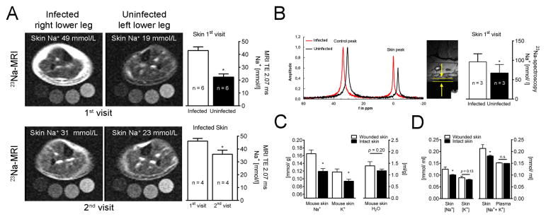
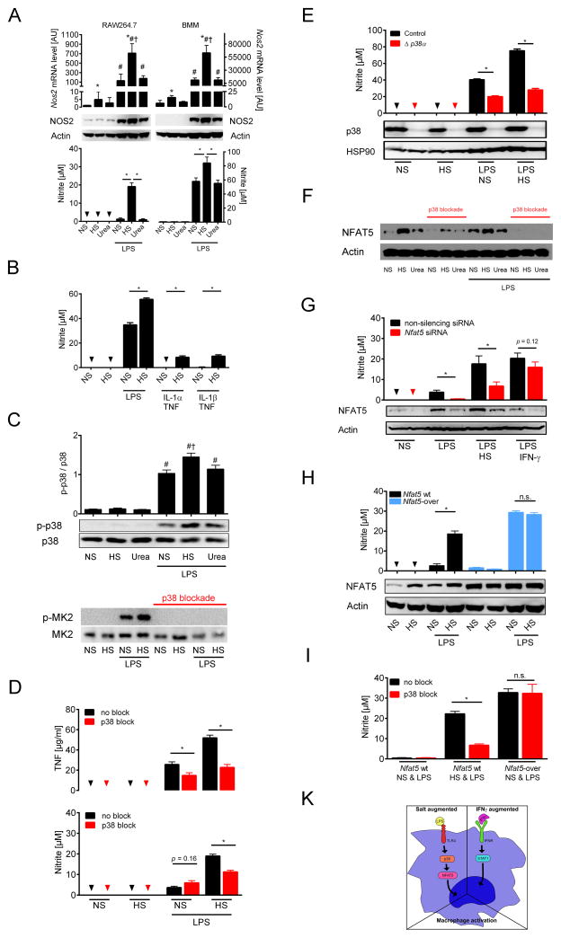
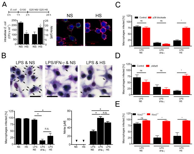
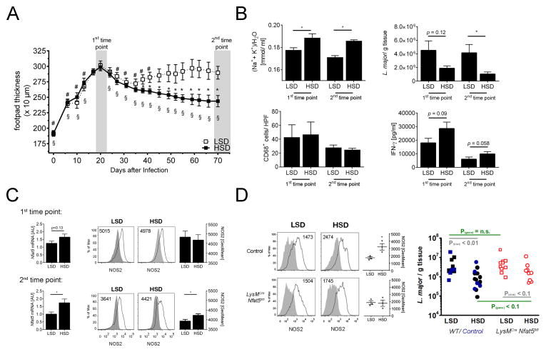
Immune cells regulate a hypertonic microenvironment in the skin; however, the biological advantage of increased skin Na(+) concentrations is unknown. We found that Na(+) accumulated at the site of bacterial skin infections in humans and in mice. We used the protozoan parasite Leishmania major as a model of skin-prone macrophage infection to test the hypothesis that skin-Na(+) storage facilitates antimicrobial host defense. Activation of macrophages in the presence of high NaCl concentrations modified epigenetic markers and enhanced p38 mitogen-activated protein kinase (p38/MAPK)-dependent nuclear factor of activated T cells 5 (NFAT5) activation. This high-salt response resulted in elevated type-2 nitric oxide synthase (Nos2)-dependent NO production and improved Leishmania major control. Finally, we found that increasing Na(+) content in the skin by a high-salt diet boosted activation of macrophages in a Nfat5-dependent manner and promoted cutaneous antimicrobial defense. We suggest that the hypertonic microenvironment could serve as a barrier to infection.


Figures

Fig. 1
Fig. 1. Infection increases Na+ storage in skin of man and mouse
(A) 23Na MRI of an infected and contralateral uninfected lower leg with bacterial skin infection. Upper left panel, acute (1st) visit; lower left panel, 28 days after antibiotics (2nd visit). Upper right panel, 23Na MRI estimates of 1st visit (mmol/l relative to standards; mean + SEM; n = 6). Lower right panel, 23Na MRI estimates (mmol/ l relative to standards) of 1st & 2nd visit (mean + SEM; n = 4). TE: echo time in ms. (B) Skin 23Na magnetic resonance spectrogram at 1st visit (skin peak). Control peak (100 mmol/l Na+ standard with shift reagent). High resolution 1H image for determination of skin thickness (arrows and bars). Skin Na+ concentrations (mean + SEM; n = 3). (C & D) Na+, K+ and water distribution in plasma and skin of animals with wounded skin (mean + SEM; n ≥ 6/group; <0.1% NaCl chow, tap water). Skin water, Na+, and K+ contents were measured. (C) Na+, K+ and H2O content per g dry weight. (D) Na+-to-water, K+-to-water and (Na++K+)-to-water ratios.
Fig. 2
Fig. 2. High salt augmented LPS-induced MΦ activation requires p38/MAPK-dependent NFAT5-signalling
(A) RAW 264.7 MΦ (left panel) and bone marrow-derived MΦ (BMM, right panel) were cultured in normal cell culture medium (NS: normal salt), with additional 40 mM NaCl in the medium (HS: high salt) or 80 mM urea ± 10 ng/ ml LPS for 24 h. Nos2 mRNA (mean + SEM; n = 4 (RAW264.7); n = 4–5 (BMM)), * P(HS) < 0.05; # P(LPS) < 0.05; † P(LPS*HS) < 0.05; NOS2 protein, and nitrite levels (mean + SEM; n = 4 (RAW264.7); n = 11 (BMM)); Triangles: not detectable (n.d.). (B) BMM were cultured in NS, with HS ± LPS (1 ng/ ml), IL-1α (50 ng/ ml) or IL-1β (50 ng/ ml) + TNF (20 ng/ ml) for 24 h. Nitrite levels (mean + SEM; 4 similar experiments); Triangles: n.d. *P < 0.05 (C) RAW 264.7 MΦ were cultured in NS, with HS or 80 mM urea ± LPS (10 ng/ ml) for 45 min. Upper panel, densitometry and immunoblotting of p38/MAPK and activated p-p38/MAPK (mean + SEM; n=8). # P(LPS) < 0.05; † P(LPS*HS) < 0.05. Lower panel, immunoblotting detected the p38/MAPK substrate MK2 and activated p-MK2. (D) RAW 264.7 MΦ were pretreated ± p38 blocker SB203580. After ½ h cells were cultured in NS, with HS ± 10 ng/ ml LPS for 24 h. Upper panel, TNF levels (mean + SEM; n = 2 in triplicates); lower panel, nitrite levels (n=7). Triangles: n.d. (E) BMM from Poly(I:C)-treated MxWT p38αfl/fl (control) and MxCre p38αfl/flp38α) mice were cultured in NS, with HS ± LPS (1 ng/ ml) for 24 h. Upper panel, nitrite levels (mean + SEM; n = 2 in quadruplicates); Triangles: n.d.; Lower panel, immunoblotting of p38/MAPK and HSP90. (F) As (D). Immunoblotting of NFAT5 and Actin. (G) RAW 264.7 MΦ electroporated with control non-silencing siRNA or Nfat5-specific siRNA (Nfat5 siRNA) were cultured in NS or HS ± LPS (10 ng/ ml) or LPS/ IFN-γ under NS for 24 h. Immunoblotting of NFAT5 and Actin. Nitrite levels (mean + SEM; n = 3–4). (H) RAW 264.7 wild-type MΦ (Nfat5 wt) and RAW 264.7 MΦ with stable Nfat5 overexpression (Nfat5-over) were cultured NS or HS ± LPS (10 ng/ ml) for 24 h. Immunoblotting of NFAT5 and Actin. Nitrite levels (mean + SEM; n = 4). (I) As (D) but in addition RAW 264.7 MΦ with stable Nfat5 overexpression (Nfat5-over) were used. A representative experiment in quintuplicates out of two independent experiments is displayed. (K) Schematic of HS-induced alterations in MΦ LPS-signaling.
Fig. 3
Fig. 3. High salt conditions promote anti-microbial activity via p38/MAPK-dependent NFAT5 signaling
(A) RAW 264.7 MΦ were infected with E. coli incubated under NS and HS conditions. Left panel, after 24 h intracellular bacterial load and nitrite levels (mean + SEM; n = 2 at least in triplicates). Right panel, after 24 h cells were fixed. GFP-E. coli, green. Phalloidin (Actin), purple. DAPI (DNA), blue. Scale bar = 20 μm. (B) BMM were infected with L. major promastigotes and stimulated with LPS (20 ng/ ml) in NS or HS medium or with IFN-γ (20 ng/ ml) under NS. Upper panel, Diff-Quik stains of BMM after 72 h. Intracellular parasites, black arrows. Scale bar = 20 μm. Lower panels, percent of infected BMM (mean + SEM; n = 5) and nitrite levels (mean + SEM; n = 7). (C) L. major-infected BMM were treated with SB203580 and stimulated as described in (A). Percent of infected BMM (mean + SEM; n = 5). (D) As (B) but BMM from Tamoxifen-treated Cre-ERT2(T)Cre Nfat5fl/flNfat5) and Cre-ERT2(T)WT/WT Nfat5fl/fl (control) mice were used (mean + SEM; n = 3). (E) As (B) but Nos2+/+ and Nos2−/− BMM were used (mean + SEM; n=3–4). Triangles: n.d.
Fig. 4
Fig. 4. High salt diet ameliorates L. major infection in vivo
(A–C) FVB WT mice were fed low salt (LSD) or high salt-diet (HSD) throughout the experiment and infected with L. major promastigotes in their footpads two weeks after initiation of the respective diet. The diets were continued throughout the experiment. (A) Lesion development of LSD and HSD mice (means ± 95% CI; n = 8/ group). * P(vs. LSD) < 0.01; # P(vs. day 20 LSD) < 0.01; § P(vs. day 20 HSD) < 0.01 (B) At the 1st time point (20–24 days after infection) and at the 2nd time point (at the end of experiment) Na+, K+ and water distribution (mean + SEM; n ≥ 6/ group), parasite burden (mean + SEM; n ≥ 7/ group), amount of lesional CD68+ MΦ (mean + SEM; n ≥ 3/ group) and Leishmania-specific T cell responses (mean + SEM; n ≥ 4/ group) are given. (C) Skin Nfat5 mRNA levels (left panel; mean + SEM; n ≥ 4) and geometric mean fluorescence of NOS2-protein expression in lesional CD11b+ cells (right panel; mean + SEM; n=4–5/ group) at the 1st time point and at the 2nd time point. Representative histograms of NOS2-expression in lesional CD11b+ cells are displayed. Insets: geometric mean fluorescence of NOS2. Grey filled area: isotype control. Black solid line: NOS2-expression. (D) LysMWT Nfat5fl/fl (control) and LysMCre Nfat5fl/fl were fed LSD and HSD and infected with L. major as described above. Left panel, as in (C) at the end of the experiment NOS2-protein expression in lesional CD11b+ cells is given (mean + SEM; n=3/ group). Right panel, WT mice (black colored), LysMCre Nfat5fl/fl (red colored) and LysMWT Nfat5fl/fl (controls, blue colored) were fed low salt (LSD) or high salt-diet (HSD) and infected with L. major promastigotes in their footpads. Parasite burden (n=9–14/ group) in skin lesions of infected mice on a HSD or LSD for over 70 days.

Comment in

References

    1. Angrisano T, Lembo F, Peluso S, Keller S, Chiariotti L, Pero R. Helicobacter pylori regulates iNOS promoter by histone modifications in human gastric epithelial cells. Med Microbiol Immunol. 2012;201:249–257. - PubMed
    1. Belkaid Y, Segre JA. Dialogue between skin microbiota and immunity. Science. 2014;346:954–959. - PubMed
    1. Braff MH, Gallo RL. Antimicrobial peptides: an essential component of the skin defensive barrier. Current topics in microbiology and immunology. 2006;306:91–110. - PubMed
    1. Buxade M, Lunazzi G, Minguillon J, Iborra S, Berga-Bolanos R, Del Val M, Aramburu J, Lopez-Rodriguez C. Gene expression induced by Toll-like receptors in macrophages requires the transcription factor NFAT5. J Exp Med. 2012;209:379–393. - PMC - PubMed
    1. Denkert C, Warskulat U, Hensel F, Haussinger D. Osmolyte strategy in human monocytes and macrophages: involvement of p38MAPK in hyperosmotic induction of betaine and myoinositol transporters. Arch Biochem Biophys. 1998;354:172–180. - PubMed

Publication types

MeSH terms

LinkOut - more resources