

Tenascin C in metastasis: A view from the invasive front
- PMID: 25738825
- PMCID: PMC4422797
- DOI: 10.1080/19336918.2015.1008331
Tenascin C in metastasis: A view from the invasive front
Abstract
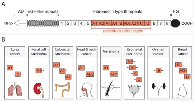
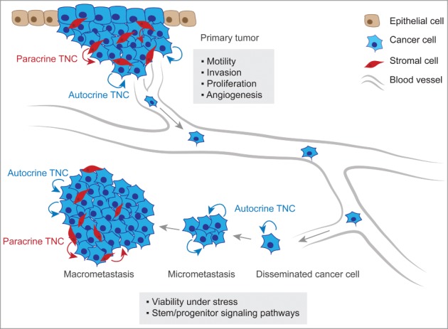
The extracellular matrix protein tenascin C (TNC) is a large glycoprotein expressed in connective tissues and stem cell niches. TNC over-expression is repeatedly observed in cancer, often at the invasive tumor front, and is associated with poor clinical outcome in several malignancies. The link between TNC expression and poor survival in cancer patients suggests a role for TNC in metastatic progression, which is responsible for the majority of cancer related deaths. Indeed, functional studies using mouse models are revealing new roles of TNC in cancer progression and underscore its important contribution to the development of metastasis. TNC has a pleiotropic role in advancing metastasis by promoting migratory and invasive cell behavior, angiogenesis and cancer cell viability under stress. TNC is an essential component of the metastatic niche and modulates stem cell signaling within the niche. This may be crucial for the fitness of disseminated cancer cells confronted with a foreign environment in secondary organs, that can exert a strong selective pressure on invading cells. TNC is a compelling example of how an extracellular matrix protein can provide a molecular context that is imperative to cancer cell fitness in metastasis.
Keywords: extracellular matrix; invasion; metastasis; niche; stem cell; tenascin C.
Figures
References
-
- Gupta GP, Massague J. Cancer metastasis: building a framework. Cell 2006; 127:679-95; PMID:17110329; http://dx.doi.org/10.1016/j.cell.2006.11.001 - DOI - PubMed
-
- Vanharanta S, Massague J. Origins of metastatic traits. Cancer cell 2013; 24:410-21; PMID:24135279; http://dx.doi.org/10.1016/j.ccr.2013.09.007 - DOI - PMC - PubMed
-
- Joyce JA, Pollard JW. Microenvironmental regulation of metastasis. Nat Rev Cancer 2009; 9:239-52; PMID:19279573; http://dx.doi.org/10.1038/nrc2618 - DOI - PMC - PubMed
-
- Bissell MJ, Hines WC. Why don't we get more cancer? A proposed role of the microenvironment in restraining cancer progression. Nat Med 2011; 17:320-9; PMID:21383745; http://dx.doi.org/10.1038/nm.2328 - DOI - PMC - PubMed
-
- Oskarsson T, Batlle E, Massague J. Metastatic stem cells: sources, niches, and vital pathways. Cell Stem Cell 2014; 14:306-21; PMID:24607405; http://dx.doi.org/10.1016/j.stem.2014.02.002 - DOI - PMC - PubMed
Publication types
MeSH terms
Substances
LinkOut - more resources
Full Text Sources
Other Literature Sources
Miscellaneous