

Development of a novel pink-eyed dilution mouse model showing progressive darkening of the eyes and coat hair with aging
- PMID: 25739360
- PMCID: PMC4427736
- DOI: 10.1538/expanim.14-0101
Development of a novel pink-eyed dilution mouse model showing progressive darkening of the eyes and coat hair with aging
Abstract
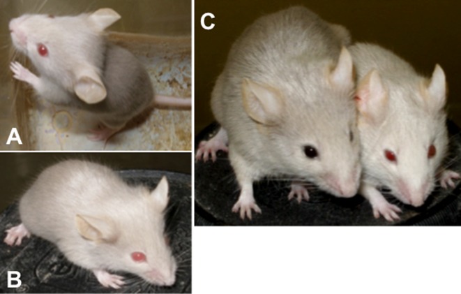
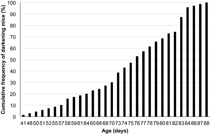
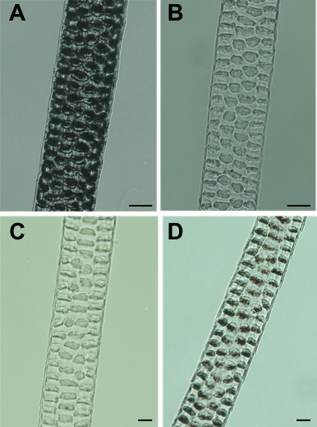
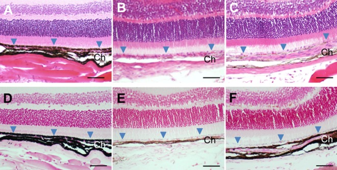
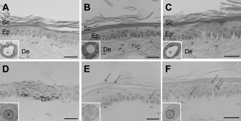
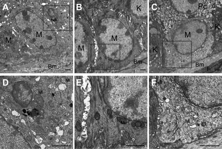
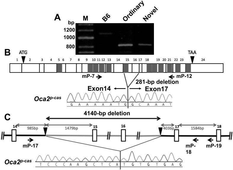
Oca2(p-cas) (oculocutaneous albinism II; pink-eyed dilution castaneus) is a coat color mutant gene on mouse chromosome 7 that arose spontaneously in wild Mus musculus castaneus mice. Mice homozygous for Oca2(p-cas) usually exhibit pink eyes and gray coat hair on the non-agouti genetic background, and this ordinary phenotype remains unchanged throughout life. During breeding of a mixed strain carrying this gene on the C57BL/6J background, we discovered a novel spontaneous mutation that causes darkening of the eyes and coat hair with aging. In this study, we developed a novel mouse model showing this unique phenotype. Gross observations revealed that the pink eyes and gray coat hair of the novel mutant young mice became progressively darker in color by approximately 3 months after birth. Light and transmission-electron microscopic observations revealed a marked increase in melanin pigmentation of coat hair shafts and choroid of the eye in the novel mice compared to that in the ordinary mice. Sequence analysis of Oca2(p-cas) revealed a 4.1-kb deletion involving exons 15 and 16 of its wild-type gene. However, there was no sequence difference between the two types of mutant mice. Mating experiments suggested that the novel mutant phenotype was not inherited in a simple fashion, due to incomplete penetrance. The novel spontaneous mutant mouse is the first example of progressive hair darkening animals and is an essential animal model for understanding of the regulation mechanisms of melanin biosynthesis with aging.
Figures

Similar articles
-
QTL Mapping for Age-Related Eye Pigmentation in the Pink-Eyed Dilution Castaneus Mutant Mouse.Genes (Basel). 2022 Jun 24;13(7):1138. doi: 10.3390/genes13071138. Genes (Basel). 2022. PMID: 35885921 Free PMC article.
-
l-tyrosine induces melanocyte differentiation in novel pink-eyed dilution castaneus mouse mutant showing age-related pigmentation.J Dermatol Sci. 2015 Dec;80(3):203-11. doi: 10.1016/j.jdermsci.2015.10.002. Epub 2015 Oct 9. J Dermatol Sci. 2015. PMID: 26475433
-
A nonsense nucleotide substitution in the oculocutaneous albinism II gene underlies the original pink-eyed dilution allele (Oca2(p)) in mice.Exp Anim. 2015;64(2):171-9. doi: 10.1538/expanim.14-0075. Epub 2015 Jan 22. Exp Anim. 2015. PMID: 25736709 Free PMC article.
-
Mouse coat colour mutations: a molecular genetic resource which spans the centuries.Bioessays. 1991 Sep;13(9):439-46. doi: 10.1002/bies.950130903. Bioessays. 1991. PMID: 1796906 Review. No abstract available.
-
The mouse p (pink-eyed dilution) and human P genes, oculocutaneous albinism type 2 (OCA2), and melanosomal pH.Pigment Cell Res. 2001 Apr;14(2):86-93. doi: 10.1034/j.1600-0749.2001.140203.x. Pigment Cell Res. 2001. PMID: 11310796 Review.
Cited by
-
QTL Mapping for Age-Related Eye Pigmentation in the Pink-Eyed Dilution Castaneus Mutant Mouse.Genes (Basel). 2022 Jun 24;13(7):1138. doi: 10.3390/genes13071138. Genes (Basel). 2022. PMID: 35885921 Free PMC article.
-
Piebaldism and chromatophore development in reptiles are linked to the tfec gene.Curr Biol. 2023 Feb 27;33(4):755-763.e3. doi: 10.1016/j.cub.2023.01.004. Epub 2023 Jan 25. Curr Biol. 2023. PMID: 36702128 Free PMC article.
-
In vitro disease modeling of oculocutaneous albinism type 1 and 2 using human induced pluripotent stem cell-derived retinal pigment epithelium.Stem Cell Reports. 2022 Jan 11;17(1):173-186. doi: 10.1016/j.stemcr.2021.11.016. Stem Cell Reports. 2022. PMID: 35021041 Free PMC article.
References
-
- Boissy R.E., Hornyak T.J.2006. Extracutaneous melanocytes. pp. 91–107. In: The Pigmentary System: Physiology and Pathophysiology, 2nd ed. (Nordlund, J.J., Boissy, R.E., Hearing, V.J., King, R.A., Oetting, W.S., and Ortonne, J.-P. eds.), Blackwell Publishing, Massachusetts.
-
- Eiberg H., Troelsen J., Nielsen M., Mikkelsen A., Mengel-From J., Kjaer K.W., Hansen L.2008. Blue eye color in humans may be caused by a perfectly associated founder mutation in a regulatory element located within the HERC2 gene inhibiting OCA2 expression. Hum. Genet. 123: 177–187. doi: 10.1007/s00439-007-0460-x - DOI - PubMed
Publication types
MeSH terms
Substances
LinkOut - more resources
Full Text Sources
Other Literature Sources
Medical
Molecular Biology Databases