. 2015 Mar 26;519(7544):431-5.
doi: 10.1038/nature14285. Epub 2015 Mar 4.

Regulated eukaryotic DNA replication origin firing with purified proteins

Affiliations

Regulated eukaryotic DNA replication origin firing with purified proteins

Joseph T P Yeeles et al. Nature. .

Abstract

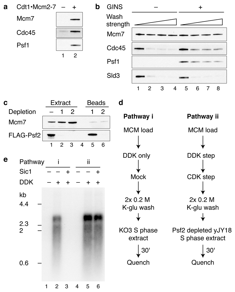
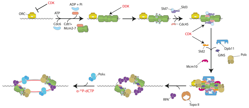
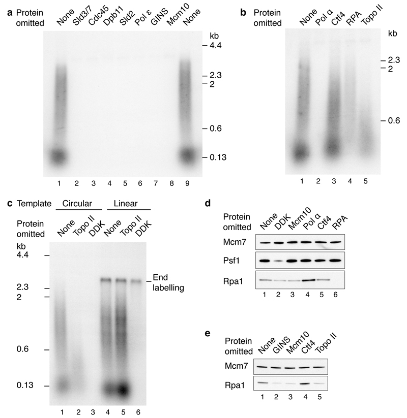
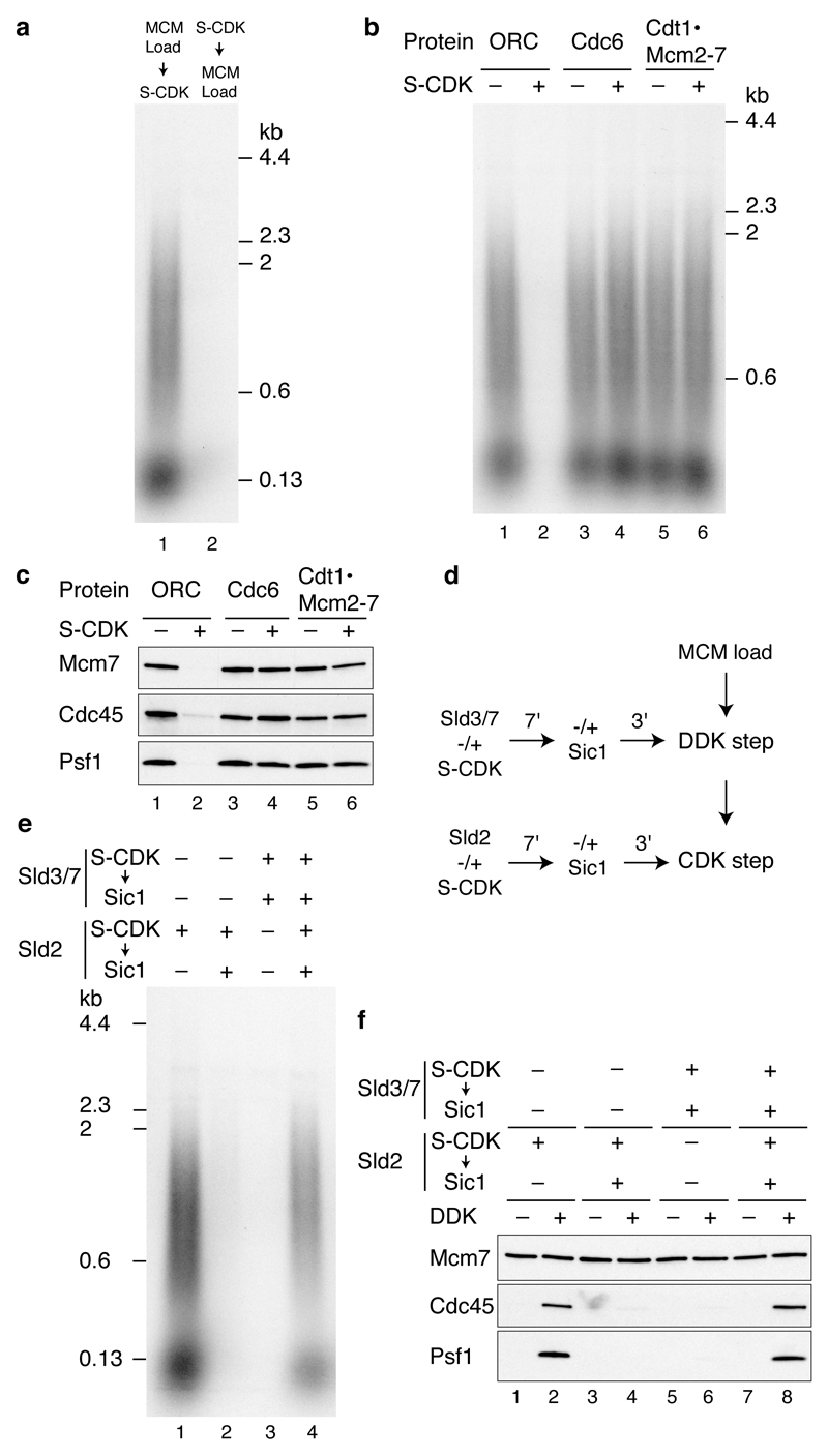
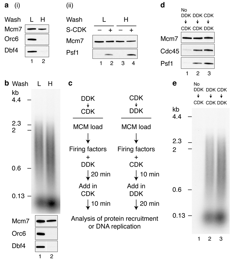
Eukaryotic cells initiate DNA replication from multiple origins, which must be tightly regulated to promote precise genome duplication in every cell cycle. To accomplish this, initiation is partitioned into two temporally discrete steps: a double hexameric minichromosome maintenance (MCM) complex is first loaded at replication origins during G1 phase, and then converted to the active CMG (Cdc45-MCM-GINS) helicase during S phase. Here we describe the reconstitution of budding yeast DNA replication initiation with 16 purified replication factors, made from 42 polypeptides. Origin-dependent initiation recapitulates regulation seen in vivo. Cyclin-dependent kinase (CDK) inhibits MCM loading by phosphorylating the origin recognition complex (ORC) and promotes CMG formation by phosphorylating Sld2 and Sld3. Dbf4-dependent kinase (DDK) promotes replication by phosphorylating MCM, and can act either before or after CDK. These experiments define the minimum complement of proteins, protein kinase substrates and co-factors required for regulated eukaryotic DNA replication.


Figures

Extended Data Figure 1
Extended Data Figure 1. Coomassie stained SDS-PAGE analysis of multisubunit complexes required for DNA replication.
a, Annotation of the polyacrylamide gels in Figs. 1a in which individual protein subunits have been labelled. b, Analysis of A-Cdk2. The protein complex consists of human Cdk2 and the bovine cyclin A-3 fragment.
Extended Data Figure 2
Extended Data Figure 2. Analysis of firing factor recruitment.
a, Immunoblots of protein recruitment conducted as in Fig. 1b but with 0.3 M KCl washes. b, Stability of recruited firing factors following washes of varying strength (lanes 1 and 5, 0.3 M K-glu; lanes 2 and 6, 0.3 M KCl; lanes 3 and 7, 0.45 M KCl; lanes 4 and 8, 0.6 M KCl). c,, Psf2-FLAG was depleted from a yJY18 S phase extract by two rounds of incubation with ANTI-FLAG M2 magnetic beads. Levels of Psf2-FLAG were determined by immunoblotting with the FLAG-M2 antibody. Soluble and bead bound protein fractions are illustrated. d, Extract-based replication reaction schemes. In pathway (i), loaded MCM is treated with DDK and added to a KO3 extract (Sld3, Sld7, Cdc45, Dpb11, Sld2 over-expression). For pathway (ii), firing factors are recruited to MCM as illustrated in Fig. 1b and the complex is added to a yJY18 extract (no firing factor over-expression) in which Psf2 (GINS complex) has been depleted. e, Replication reactions as described in d using A-Cdk2 for firing factor recruitment. Where indicated, Sic1 was added to the extract 20 min prior to replication.
Extended Data Figure 3
Extended Data Figure 3. Characterisation of the in vitro DNA replication reaction.
a, b, Replication reactions conducted as in Fig. 3a with A-Cdk2 on ARS1 circular DNA templates for 1 hour. c, Time course of a standard replication reaction using A-Cdk2 on the ARS1 linear DNA template. d, Quantitation of a time course conducted as in c. DNA synthesis was normalised to the total DNA synthesis at 90 minutes. Small and large replication products were not quantified separately at 5 min as they are not well resolved at this time point. e, Pulse chase experiment conducted with A-Cdk2 on the ARS1 linear DNA template. For the pulse the dCTP concentration was reduced to 4 μM. Following a 10 min incubation unlabelled dCTP was added to 100 μM.
Extended Data Figure 4
Extended Data Figure 4. Characterisation of RPA recruitment.
Unless stated reactions were conducted on ARS1 circular templates. a, Vaccinia virus topoisomerase I supports DNA replication with purified proteins. Replication reactions with either Topo II (25 nM) or Vaccinia virus topoisomerase I (0.125 units/μl). Two different Topo II fractions (Fr1 and Fr2) were used for comparison. b, Nucleotide dependence of RPA recruitment in a complete replication reaction with Topo II. c, RPA recruitment reactions were conducted on ARS1 circular template in the presence of Vaccinia virus topoisomerase I (0.125 units/μl), or on a linear template in the absence of a topoisomerase. dNTPs, C/G/UTP, pol α and Ctf4 were omitted from the final step of the reaction.
Extended Data Figure 5
Extended Data Figure 5. Regulation of MCM loading and origin firing by S-CDK is ATP dependent.
a, Replication reaction where ORC was pre-incubated with S-CDK prior to MCM loading. When Sic1 was added before ORC the mix was incubated for 5 min and ORC was then added for 10 min. b, Pre-incubation of ORC with S-CDK in the presence or absence of ATP. After incubation with Sic1, ATP was added to the reaction lacking ATP. c, Sld3/7 and Sld2 were pre-incubated with S-CDK as illustrated in Fig. 5d in the presence or absence of ATP. Following incubation with Sic1, samples that did not contain ATP for the pre-incubation step were supplemented with ATP.
Extended Data Figure 6
Extended Data Figure 6. Cartoon illustrating protein kinase regulated eukaryotic DNA replication origin firing with purified proteins.
Firing factors are recruited to loaded MCM in a DDK- and CDK-dependent manner. DNA synthesis is initiated once the DNA template has been unwound. CDK also functions to inhibit MCM loading by phosphorylating ORC.
Extended Data Figure 7
Extended Data Figure 7. Internally FLAG-tagged Cdc45 supports normal DNA replication in S phase extracts.
a, The previously reported interaction between Sld3 and Cdc45 was exploited to co-immunoprecipitate Cdc45 from yJY16 extracts (Dpb11 and Sld2 over-expression) by incubation with FLAG-Sld3/7 that was pre-coupled to ANTI-FLAG M2 magnetic beads. b, In vitro extract-based replication reaction on soluble ARS1 circular template using yJY16 extracts where endogenous Cdc45 was depleted as indicated. The extract was supplemented with purified Sld3/7 as the complex is not over-expressed in yJY16. The experiment was conducted for 30 min as described previously and products were separated through a 1% native agarose gel. Internally FLAG-tagged Cdc45 (52 nM) was added back as indicated. The locations of the different replication products are illustrated.
Figure 1
Figure 1. DDK- and CDK-dependent firing-factor recruitment with purified proteins.
a, Purified MCM loading and firing factors (i), and additional factors required for DNA replication (ii) analysed by SDS-PAGE with Coomassie staining. b, Reaction scheme for firing-factor recruitment. c, d, Immunoblots of recruitment reactions conducted as illustrated in bon ARS305 linear DNA.
Figure 2
Figure 2. The purified firing factors are functional.
a, b, Immunoblots of recruitment reactions performed as in Fig. 1b using ARS305 linear DNA with either, a, 0.3 M K-glutamate (K-glu) or b, 0.3 M KCl washes. c, Scheme for extract-based replication reactions. Unless otherwise stated, in this and all subsequent experiments, components of the ‘CDK’ and ‘DDK’ steps are as in Fig. 1. Firing factors were recruited as illustrated in Fig. 1b. Beads were isolated, washed twice and added to an S phase extract not overexpressing firing factors (yJY18) and where the Psf2 subunit of the GINS complex was immunodepleted (Extended Data Fig. 2c). d, Reaction performed as in c. Nascent DNA was labelled by including α32P-dCTP in the extract step and products were separated through a 0.6% alkaline agarose gel.
Figure 3
Figure 3. The initiation of DNA synthesis with purified proteins.
a, Reaction pathway for DNA replication with purified proteins. Firing factors were bound to MCM, the complex isolated, and added to a new buffer containing proteins required for DNA synthesis (Fig. 1a (i)). b, Replication reaction conducted as shown in a on ARS1 circular DNA. In this and all subsequent replication assays products were separated through 0.7% alkaline agarose gels. c, Replication on ARS1 linear templates. (i) Photocleaved DNA templates analysed by native agarose gel electrophoresis and ethidium bromide staining. (ii) Replication following the pathway shown in a. The MCM loading conditions were modified to confer origin specificity on the replication reaction (see Methods for details).
Figure 4
Figure 4. Requirements for origin firing.
Reactions were performed as illustrated in Fig. 3a on circular templates unless stated. a, Firing factors required to initiate DNA synthesis. b, Protein dependencies of DNA replication for components functioning downstream of firing factor recruitment. c, Topoisomerase dependence on circular and linear ARS1 templates. d, RPA recruitment in a complete replication reaction. e, Dependence of RPA recruitment in the absence of pol α.
Figure 5
Figure 5. Regulation of MCM loading and origin firing by S-CDK.
a,Replication reactions where MCM loading factors were incubated with S-CDK before or after MCM loading on ARS1 circular DNA. Replication, b, and protein recruitment, c, on ARS1 circular DNA where ORC, Cdc6 and Cdt1·Mcm2-7 were individually incubated with S-CDK followed by addition of Sic1. The remaining MCM loading factors were added and reactions performed as shown in Fig. 3a (replication) and Fig. 1b (recruitment). d, Scheme to phosphorylate Sld3/7 and Sld2 in isolation from MCM and firing factors. Prior to both the DDK step and CDK step Sld3/7 and Sld2 were incubated with S-CDK before addition of Sic1. The remaining firing factors were added together with isolated DNA beads and each reaction step was performed as for standard reactions. e, Replication reaction staged as illustrated in d. f, Firing factor recruitment conducted as in d, followed by 0.3 M KCl washes.
Figure 6
Figure 6. DDK phosphorylation of MCM promotes origin firing either before or after S-CDK.
a, Reactions performed essentially as in Fig. 1b on circular DNA but with an additional mid-reaction wash immediately following DDK phosphorylation, in either a low salt (L) (0.3 M K-glu), or high salt (H) (0.6 M NaCl) buffer. Bound proteins immediately following the mid-reaction wash (i), and after firing factor recruitment followed by 0.3 M KCl washes (ii). b, Replication reactions with either high or low salt mid-reaction washes. c, Reaction schemes to test the order of DDK and CDK action. Firing factor recruitment was performed in a single stepwith DDK and CDK added at different times as indicated. Protein recruitment, d, and replication, e, performed with S-CDK as illustrated in c.

Comment in

References

    1. Tanaka S, Araki H. Helicase Activation and Establishment of Replication Forks at Chromosomal Origins of Replication. Cold Spring Harbor perspectives in biology. 2013 - PMC - PubMed
    1. Siddiqui K, On KF, Diffley JFX. Regulating DNA Replication in Eukarya. Cold Spring Harbor perspectives in biology. 2013 - PMC - PubMed
    1. Remus D, et al. Concerted loading of Mcm2-7 double hexamers around DNA during DNA replication origin licensing. Cell. 2009;139:719–730. - PMC - PubMed
    1. Evrin C, et al. A double-hexameric MCM2-7 complex is loaded onto origin DNA during licensing of eukaryotic DNA replication. Proc Natl Acad Sci U S A. 2009;106:20240–20245. - PMC - PubMed
    1. Gros J, Devbhandari S, Remus D. Origin plasticity during budding yeast DNA replication in vitro. EMBO J. 2014;33:621–636. - PMC - PubMed

Publication types

MeSH terms