Review
. 2015 Aug;43(6):776-93.
doi: 10.1177/0192623315570339. Epub 2015 Mar 3.

Vaginal Cytology of the Laboratory Rat and Mouse: Review and Criteria for the Staging of the Estrous Cycle Using Stained Vaginal Smears

Affiliations
Review

Vaginal Cytology of the Laboratory Rat and Mouse: Review and Criteria for the Staging of the Estrous Cycle Using Stained Vaginal Smears

Michelle C Cora et al. Toxicol Pathol. 2015 Aug.

Abstract

Microscopic evaluation of the types of cells present in vaginal smears has long been used to document the stages of the estrous cycle in laboratory rats and mice and as an index of the functional status of the hypothalamic-pituitary-ovarian axis. The estrous cycle is generally divided into the four stages of proestrus, estrus, metestrus, and diestrus. On cytological evaluation, these stages are defined by the absence, presence, or proportion of 4 basic cell types as well as by the cell density and arrangement of the cells on the slide. Multiple references regarding the cytology of the rat and mouse estrous cycle are available. Many contemporary references and studies, however, have relatively abbreviated definitions of the stages, are in reference to direct wet mount preparations, or lack comprehensive illustrations. This has led to ambiguity and, in some cases, a loss of appreciation for the encountered nuances of dividing a steadily moving cycle into 4 stages. The aim of this review is to provide a detailed description, discussion, and illustration of vaginal cytology of the rat and mouse estrous cycle as it appears on smears stained with metachromatic stains.

Keywords: diestrus; estrus; metestrus.; proestrus.


Figures

Figure 1.
Figure 1.
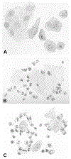
Historical representation of select stages of the rat estrous cycle as presented in Long and Evans (1922). These eloquent pencil drawings illustrate proestrus (A), metestrus (B) and diestrus (C).
Figure 2.
Figure 2.
Example of a commercially-available 1” x 3” glass slide with a pre-drawn numbered grid for vaginal cytology sample collection. This slide contains samples that were collected daily for eight consecutive days from an individual animal and, after dry fixation, were stained with a metachromatic stain.
Figure 3.
Figure 3.
Many neutrophils (arrows), characterized by their very small size and multilobulated nuclei, and two large nucleated epithelial cells (arrowhead) are present on this diestrous vaginal smear from a Sprague Dawley rat. Toluidine blue O stain. In Elmore et el (20XX) Proceedings of the 2014 National Toxicology Program satellite symposium, Toxicol Pathol, TBD.
Figure 4.
Figure 4.
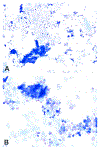
Ruptured neutrophils (arrows) are present on these vaginal cytology smears from a Sprague Dawley rat (A) and B6C3F1/N mouse (B). Toluidine blue O stain. Original objective magnification of 20X (B) or 40X (A).
Figure 5.
Figure 5.
Small nucleated epithelial cells from a proestrous vaginal smear of a Sprague Dawley rat. Occasional nucleated epithelial cells containing small cytoplasmic vacuoles (arrow) and a few anucleated epithelial cells are also present. Toluidine blue O stain. Original objective magnification of 40X.
Figure 6.
Figure 6.
Several large nucleated epithelial cells (arrows) are intermixed with anucleated epithelial cells (arrowheads) in a vaginal smear of late estrus from a Sprague Dawley rat. Numerous bacteria are observed adhered to many of the epithelial cells. Modified Wright – Giemsa stain. Original objective magnification of 40X. In Elmore et el (20XX) Proceedings of the 2014 National Toxicology Program satellite symposium, Toxicol Pathol, TBD.
Figure 7.
Figure 7.
A large nucleated epithelial cell containing many small cytoplasmic granules (arrow). Sprague Dawley rat. Modified Wright – Giemsa stain. Original oil objective magnification of 100X.
Figure 8.
Figure 8.
Anucleated epithelial cells from a vaginal smear of estrus in a B6C3F1/N mouse. Some cells possess ghost nuclei (arrows). Modified Wright – Giemsa stain. Original objective magnification of 40X. In Elmore et el (20XX) Proceedings of the 2014 National Toxicology Program satellite symposium, Toxicol Pathol, TBD.
Figure 9.
Figure 9.
A keratin bar (arrow) is observed in a vaginal smear of estrus from a Sprague Dawley rat. Note that many bacteria are either adhered to the cells or free in the background. Modified Wright – Giemsa stain. Original objective magnification of 40X.
Figure 10.
Figure 10.
Estrous cycle wheels. Visual representation of the cell types and relative proportion of each cell type present during the four stages of the estrous cycle in the mouse and rat. The size of each quadrant does not directly correlate to the length of each stage. Adapted from: Byers, S. L., Wiles, M. V., Dunn, S.L., and Taft, R. A. (2012). Mouse estrous cycle identification tool and images. Plos One 7, 1-5. DOI: 10.1371/journal.pone.0035538. The mouse wheel was modified from its original form to create the rat wheel. Adaptations and modifications by David Sabio.
Figure 11.
Figure 11.
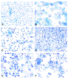
Vaginal smears of proestrus from Sprague-Dawley rats characterized by high numbers of small nucleated epithelial cells found individually and in cohesive clusters (arrows). Proestrus appears the same in mice. Plate D shows the appearance of a so-called epithelial “strand” that is typical of proestrus. Modified Wright – Giemsa stain (A, D) and Toluidine blue O stain (B, C). Original objective magnification of 10X (A, B), 20X (C) or 40X (D). Plate C is in Elmore et el (20XX) Proceedings of the 2014 National Toxicology Program satellite symposium, Toxicol Pathol, TBD.
Figure 12.
Figure 12.
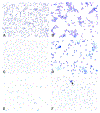
Representative photomicrographs of the first (A, C) and second (B, D) “phases” of estrus in mice. The first phase typically has smaller anuclear epithelial cells that are arranged in loose clusters reminiscent of proestrus. The anuclear epithelial cells of the second phase are generally larger, more evenly dispersed and higher in numbers. Plates C and D are two consecutive days of estrus from the same B6C3F1/N mouse. Modified Wright – Giemsa stain (A, B) and Toluidine blue O stain (C, D). Original objective magnification of 10X.
Figure 13.
Figure 13.
Vaginal smears of estrus (A, B) and late estrus (C – F) in several Sprague Dawley rats. Plates A and B represent a typical estrus stage with high numbers of anucleated epithelial cells. Vaginal smears of the late estrus phase (C – F) are characterized by the presence of round, oval and spindle shaped nucleated epithelial cells interspersed among anucleated epithelial cells. Modified Wright – Giemsa stain (A – C, E) and Toluidine blue O stain (D, F). Original objective magnification of 10X (A, C, D), 20X (F) or 40X (B, E). Plate A is in Elmore et el (20XX) Proceedings of the 2014 National Toxicology Program satellite symposium, Toxicol Pathol, TBD.
Figure 14.
Figure 14.
Metestrous smears from several B6C3F1/N mice. Metestrus begins with the emergence of neutrophils interspersed or clumped (arrow) among the anucleated epithelial cells (A – D). Occasional nucleated epithelial cells may also be present. As metestrus progresses, neutrophil numbers increase resulting in smears of very high cellularity (E, F). Neutrophil and epithelial cell numbers decrease as the mouse transitions to diestrus (G, H). Plates A and D illustrate two consecutive days from a control B6C3F1/N mouse, representing the uncommon occurrence of a metestrus stage being recorded for 2 days in a row in a mouse. Modified Wright – Giemsa stain (A – D, G, H) and Toluidine blue O stain (E, F). Original objective magnification of 10X (A, C – E, G), 20X (F, H), or 40X (B). Plate F is in Elmore et el (20XX) Proceedings of the 2014 National Toxicology Program satellite symposium, Toxicol Pathol, TBD.
Figure 14.
Figure 14.
Metestrous smears from several B6C3F1/N mice. Metestrus begins with the emergence of neutrophils interspersed or clumped (arrow) among the anucleated epithelial cells (A – D). Occasional nucleated epithelial cells may also be present. As metestrus progresses, neutrophil numbers increase resulting in smears of very high cellularity (E, F). Neutrophil and epithelial cell numbers decrease as the mouse transitions to diestrus (G, H). Plates A and D illustrate two consecutive days from a control B6C3F1/N mouse, representing the uncommon occurrence of a metestrus stage being recorded for 2 days in a row in a mouse. Modified Wright – Giemsa stain (A – D, G, H) and Toluidine blue O stain (E, F). Original objective magnification of 10X (A, C – E, G), 20X (F, H), or 40X (B). Plate F is in Elmore et el (20XX) Proceedings of the 2014 National Toxicology Program satellite symposium, Toxicol Pathol, TBD.
Figure 15.
Figure 15.
Metestrous vaginal smears from several Sprague Dawley rats. Metestrus begins with the emergence of neutrophils interspersed or clumped among the nucleated and anucleated epithelial cells (A, B). Note that the appearance of metestrus differs in rats and mice, as rats have much higher numbers of nucleated epithelial cells. As metestrus progresses, neutrophil numbers increase resulting in smears of high cellularity (C - F). Toluidine blue O stain. Original objective magnification of 10X (A, C, E) or 40X (B, D, F).
Figure 16.
Figure 16.
Diestrous vaginal smears from B6C3F1/N mice (A – E) and a Sprague Dawley rat (F) characterized by a moderate to low cellularity with predominately neutrophils that are mixed with lower numbers of epithelial cells. A small clump of round epithelial cells (F; arrow) is present on a diestrous smear taken the day before the emergence of proestrus. Modified Wright – Giemsa stain (C – E) and Toluidine blue O stain (A, B, F). Original objective magnification of 10X (A, C, E, F) or 40X (B, D). Plate E is in Elmore et el (20XX) Proceedings of the 2014 National Toxicology Program satellite symposium, Toxicol Pathol, TBD.
Figure 17.
Figure 17.
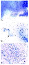
Diestrous vaginal smears from several Sprague Dawley rats. Basophilic to eosinophilic staining mucous has entrapped and distorted the epithelial cells and neutrophils. A classic mucous “swirl” is seen in plate B (arrow). Modified Wright – Giemsa stain (B) and Toluidine blue O stain (A, C).
Figure 18.
Figure 18.
Lower (Original objective magnification of 10X) and higher (20X) magnifications of a vaginal cytology smear taken from a Sprague Dawley rat representing a proestrus-estrus transition. Individual and clumps of small epithelial cells are seen among anucleated epithelial cells. Based on complete review of this smear, the recorded stage (based on the four stage paradigm) was proestrus or “P”. Using the other classification paradigm discussed in the article it could also be classified as “P(e)”. Please refer to the section on transitional stages for further details. Modified Wright – Giemsa stain.

References

    1. Allen E The oestrous cycle in the mouse. (1922) Am J Anat 30, 297–371.
    1. Astwood EB (1939). Changes in the weight and water content of the uterus of the normal adult rat. Am J Physiol 126, 162–70.
    1. Blandau RJ, Boling JL, and Young WC (1941). The length of heat in the albino rat as determined by copulatory response. Anat Rec 79, 453–63.
    1. Butcher RL, Collins WE, and Fugo NW (1974). Plasma concentration of LH, FSH, prolactin, progesterone and estradiol-17β throughout the 4-day estrous cycle of the rat. Endocrinology 94, 1704–8. - PubMed
    1. Byers SL, Wiles MV, Dunn SL, and Taft RA (2012). Mouse estrous cycle identification tool and images. Plos One 7, 1–5. - PMC - PubMed

Substances

LinkOut - more resources