. 2015 Mar 4;7(277):277ra28.
doi: 10.1126/scitranslmed.aaa1405.

Liver-directed lentiviral gene therapy in a dog model of hemophilia B

Affiliations

Liver-directed lentiviral gene therapy in a dog model of hemophilia B

Alessio Cantore et al. Sci Transl Med. .

Abstract

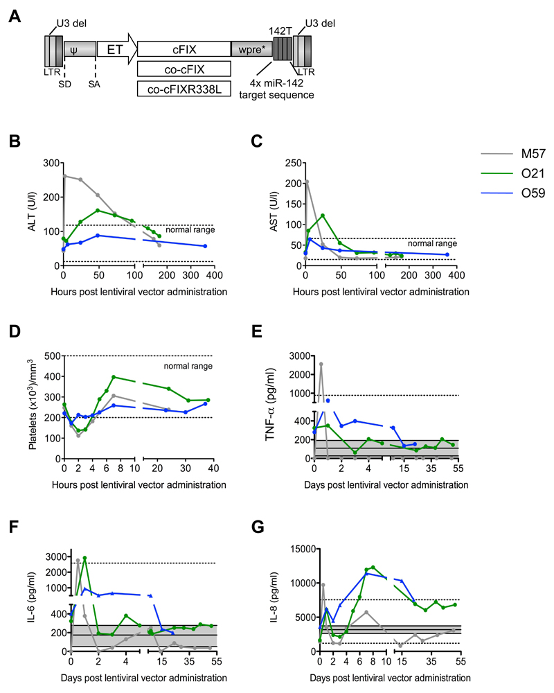
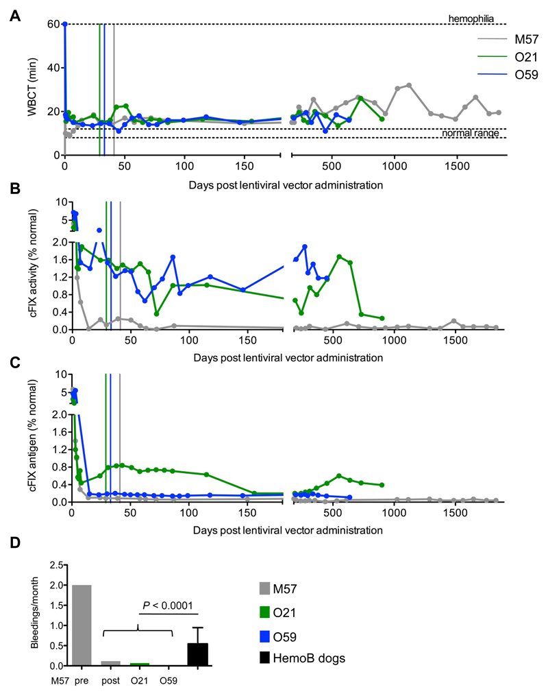
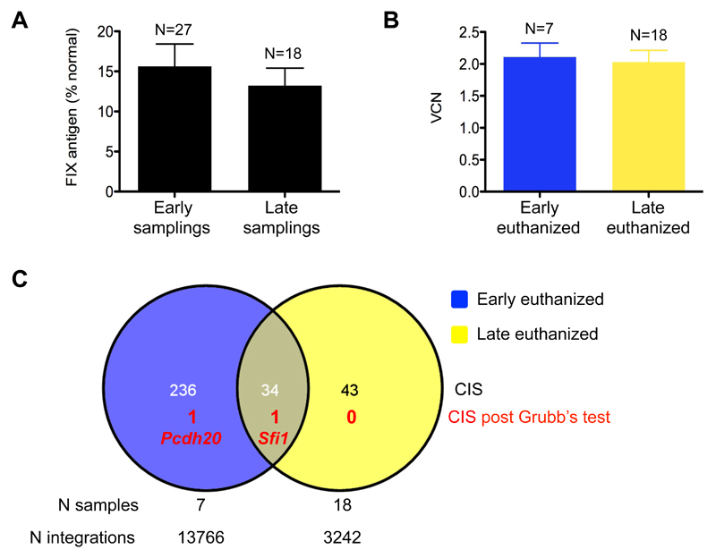
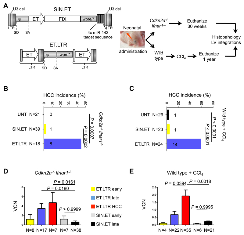
We investigated the efficacy of liver-directed gene therapy using lentiviral vectors in a large animal model of hemophilia B and evaluated the risk of insertional mutagenesis in tumor-prone mouse models. We showed that gene therapy using lentiviral vectors targeting the expression of a canine factor IX transgene in hepatocytes was well tolerated and provided a stable long-term production of coagulation factor IX in dogs with hemophilia B. By exploiting three different mouse models designed to amplify the consequences of insertional mutagenesis, we showed that no genotoxicity was detected with these lentiviral vectors. Our findings suggest that lentiviral vectors may be an attractive candidate for gene therapy targeted to the liver and may be potentially useful for the treatment of hemophilia.


Conflict of interest statement

Competing interests: LN is an inventor on pending and issued patents on lentiviral vector technology and miR-regulated lentiviral vectors (Gene Vector, WO2007000668).

Figures

Figure 1
Figure 1. Intraportal administration of lentiviral vectors to dogs with hemophilia B.
(A) Schematic representation of the third-generation Self Inactivating (SIN) lentiviral vectors (proviral form) used in this work [U3 del: deletion of the promoter/enhancer of the HIV Long Terminal Repeats (LTR) (43)]. SD: splicing donor site. SA: splicing acceptor site. ψ: packaging signal. Wpre*: mutated woodchuck hepatitis virus post-transcriptional regulatory element (44). 142T: miR-142 target sequence made of 4 tandem copies of a sequence perfectly complementary to miR-142. Hepatocyte-specific Enhanced Transthyretin (ET) promoter composed of synthetic hepatocyte-specific enhancers and transthyretin promoter (45). The wildtype, codon-usage optimized, and codon-usage optimized and hyper-functional cDNAs of canine factor IX (cFIX, co-cFIX, co-cFIXR338L) were used as transgenes (14). Serum concentrations of alanine aminotransferase, ALT (B) and aspartate aminotransferase, AST (C), platelet counts (D), and serum concentrations of TNF-α (E), IL-6 (F) and IL-8 (G) were measured in blood samples collected at the indicated time points after lentiviral vector administration to dogs M57 (grey line), O21 (green line), and O59 (blue line). Baseline values are shown as “time 0”. (B-D) The normal range is shown (dashed lines). (E-G) The mean ± standard deviation (grey area) and range (dashed lines) of the serum concentrations of each cytokine measured in samples collected from 11 control untreated dogs are shown. Note that the lowest range for TNF-α and IL-6 is 0. Dog O59 was administered corticosteroids and anti-histamine drugs before lentiviral vector infusion to reduce inflammation.
Figure 2
Figure 2. Lentiviral vector-mediated gene therapy targeted to liver provides stable improvement in clotting time in dogs with hemophilia B.
Whole blood clotting time, WBCT (A) measured in blood samples, canine factor IX activity (cFIX) (B) and cFIX antigen (C) measured by activated partial thromboplastin time, aPTT (B) or ELISA (C) in plasma samples collected at the indicated times after lentiviral vector administration from dogs M57 (grey line), O21 (green line), O59 (blue line) 0. The colored vertical lines indicate 27 days after the last normal plasma transfusion of the dogs at which time exogenous canine factor IX had been washed-out. (D) Frequency of spontaneous bleedings (bleeding events/month of observation) in the treated dogs after gene therapy. For M57, the frequency of spontaneous bleeding before gene therapy is shown. The mean ± SD bleeding frequency of 10 untreated dogs with hemophilia B in the colony is shown (black bar) (46). P < 0.0001 (2-sample test for equality of proportions; see also table S6).
Fig. 3
Fig. 3. No evidence of genotoxicity after lentiviral vector integration into liver of mice.
(A) Factor IX antigen measured by ELISA in the plasma collected from mice “early” (<3 months) or “later” (6-12 months) after lentiviral vector administration. P = 0.391, Student’s t test. (B) Vector copy number (VCN) in liver DNA collected from mice euthanized early or later after lentiviral vector administration. P = 0.806, Student’s t test. (A, B) Data are mean±standard error of the mean (SEM). (C) Venn diagram representing Common Insertion Sites (CIS) identified in liver DNA of mice euthanized early or later after lentiviral vector administration. The overlap is calculated considering the gene associated with each CIS; the number of CIS that passed the Grubb’s test is shown along with the gene name. The number of samples analyzed and integration sites retrieved are indicated for the two data sets.
Figure 4
Figure 4. SIN.ET lentiviral vectors do not induce hepatocellular carcinoma in tumor-prone mice.
(A) Experimental outline of the in vivo biosafety study in mice. (Left) Schematic representations of the lentiviral vectors used U3 del: deletion of the promoter/enhancer of the HIV Long Terminal Repeats (LTR) (43). SD: splicing donor site. SA: splicing acceptor site. ψ: packaging signal. Wpre*: mutated woodchuck hepatitis virus post-transcriptional regulatory element (44). 142T: miR-142 target sequence made of 4 tandem copies of a sequence perfectly complementary to miR-142. Hepatocyte-specific Enhanced Transthyretin (ET) promoter composed of synthetic hepatocyte-specific enhancers and transthyretin promoter (45). Either SIN.ET (gene therapy lentiviral vector with Self Inactivating Long Terminal Repeats and an internal Enhanced Transthyretin promoter) or ET.LTR (oncogenic lentiviral vector with transcriptionally active Long Terminal Repeats containing the Enhanced Transthyretin promoter) were administered at matched doses to newborn Cdkn2a-/-Ifnar1-/- tumor-prone mice or wildtype mice resulting in four experimental groups. Wildtype mice then were given a CCl4-based tumor promoting regimen. Mice were euthanized at the indicated time points or earlier if sick. Necropsy was performed and samples were collected for DNA extraction (for determination of vector copy number and the retrieval of integration sites ) and for histopathology analysis. (B, C) Shown is the incidence of hepatocellular carcinoma (HCC) in Cdkn2a-/-Ifnar1-/- mice (B) or wildtype mice (C) transduced with the two different lentiviral vectors (SIN.ET or ET.LTR) or untransduced (UNT). Untransduced mice include historical controls (n=20 Cdkn2a-/-Ifnar1-/- and n=9 wildtype mice) (27). P values were calculated by two-tailed Fisher’s exact test. Numbers on the histograms indicate the number of mice that developed HCC. (D, E) Vector copy number in liver DNA from Cdkn2a-/-Ifnar1-/- mice (D) or wildtype mice (E) collected two weeks after lentiviral vector administration (early) or at necropsy (late). Data are mean±SEM. P values were calculated by One-Way ANOVA and Bonferroni’s multiple correction test. All vector copy numbers were measured in non-tumoral liver tissue except for ET.LTR-induced HCCs.
Fig. 5
Fig. 5. Integration sites analysis does not reveal genotoxicity of SIN.ET lentiviral vectors in tumor-prone mice.
(A) Venn diagram representing Common Insertion Sites (CIS) identified in liver DNA of SIN.ET-transduced and ET.LTR-transduced mice. The overlap is calculated considering the gene associated with each CIS. The number of CIS that passed the Grubb’s test is shown with the gene name (red). The number of samples analyzed and the total number of integration sites are indicated for the two data sets. (B, C) Schematic drawing of two representative CIS of ET.LTR (B) and SIN.ET (C) lentiviral vectors. Each colored bar represents an integration site (red: from ET.LTR-induced HCCs; orange: from non-tumoral liver of mice transduced with ET.LTR; black: from liver of mice transduced with SIN.ET). Colored arrows indicate the orientation of the integration site. The gene within the region is represented below, with black boxes indicating exons and arrows indicating the transcription orientation. The span of the outlined genomic region is indicated on top. (D) Common insertion sites (CIS) power, calculated as the number of different integration sites targeting each CIS. (E) CIS representation, calculated as % of sequencing reads from all integration sites comprised within a CIS over the total number of reads within an experimental data set. (D, E) Data are mean±SEM. P values were calculated by One-Way ANOVA and Bonferroni’s multiple correction test. For all the analyses, integration sites from the two mouse models were merged.

References

    1. Bolton-Maggs PH, Pasi KJ. Haemophilias A and B. Lancet. 2003;361:1801–1809. - PubMed
    1. High KH, Nathwani A, Spencer T, Lillicrap D. Current status of haemophilia gene therapy. Haemophilia. 2014;20(Suppl 4):43–49. - PubMed
    1. Berntorp E, Shapiro AD. Modern haemophilia care. Lancet. 2012;379:1447–1456. - PubMed
    1. Astermark J, Santagostino E, Keith Hoots W. Clinical issues in inhibitors. Haemophilia. 2010;16(Suppl 5):54–60. - PubMed
    1. Franchini M, Mannucci PM. Past, present and future of hemophilia: a narrative review. Orphanet J Rare Dis. 2012;7:24. - PMC - PubMed

Publication types