. 2015 Apr 3;14(4):1920-36.
doi: 10.1021/pr5013015. Epub 2015 Mar 18.

Host-pathogen interaction profiling using self-assembling human protein arrays

Affiliations

Host-pathogen interaction profiling using self-assembling human protein arrays

Xiaobo Yu et al. J Proteome Res. .

Abstract

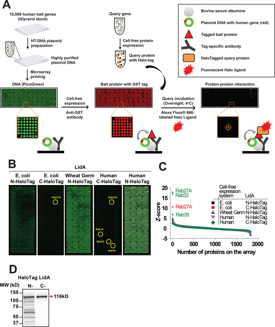
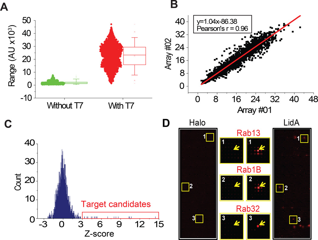
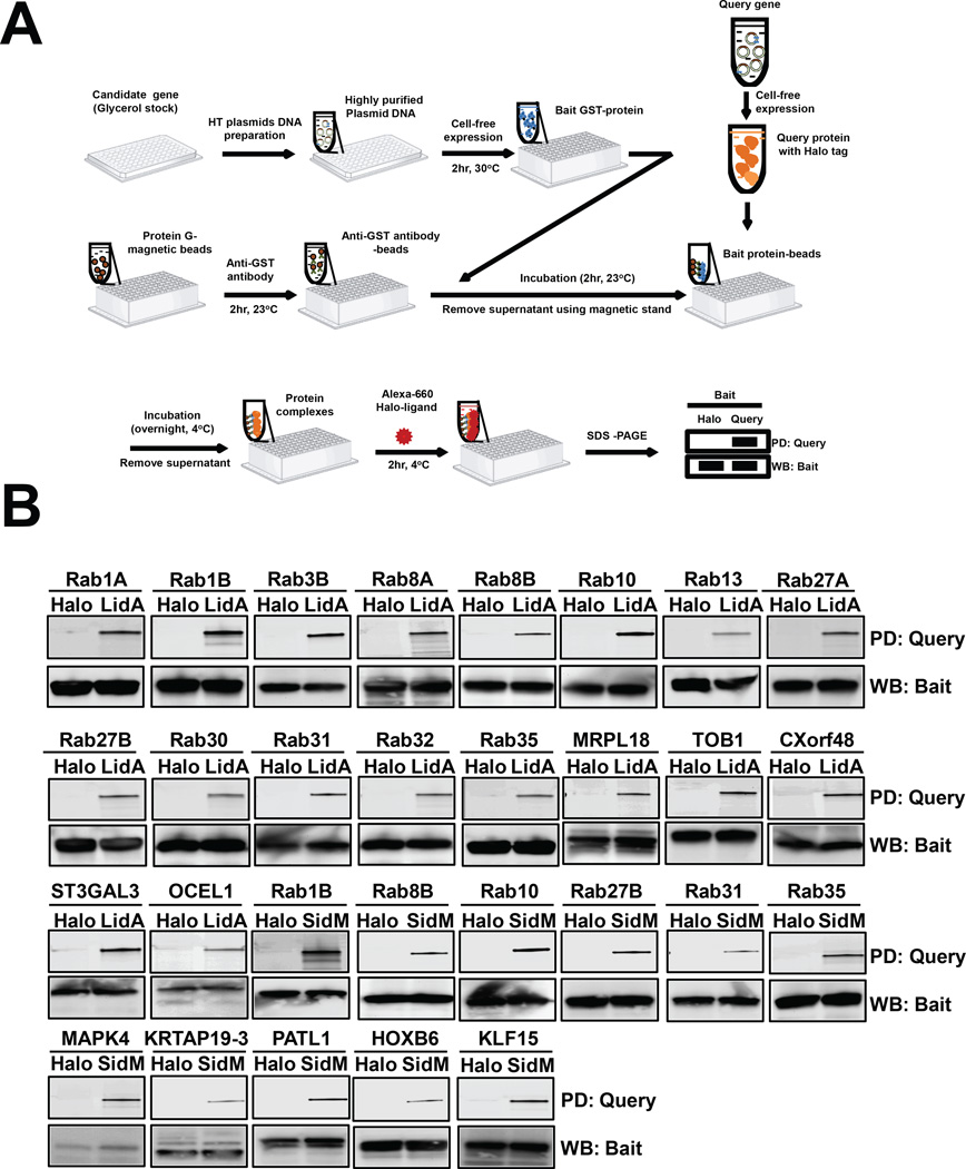
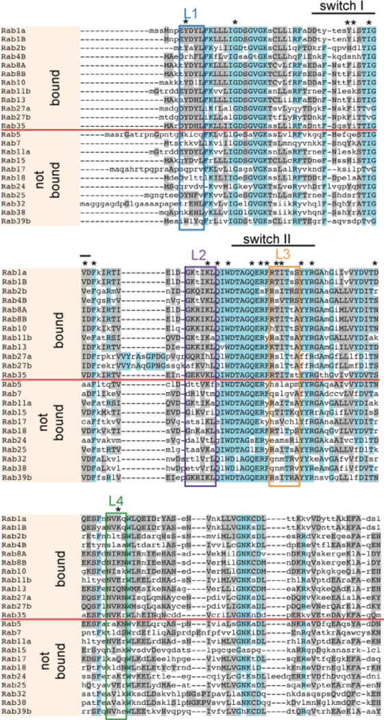
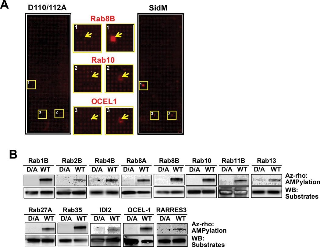
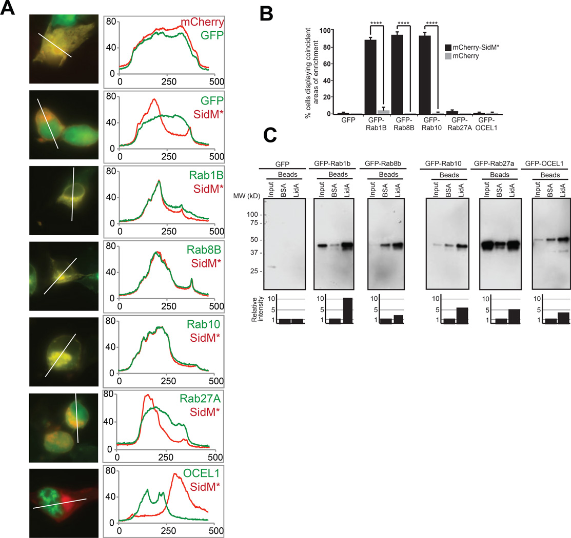
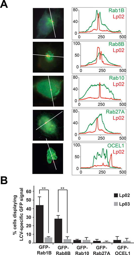
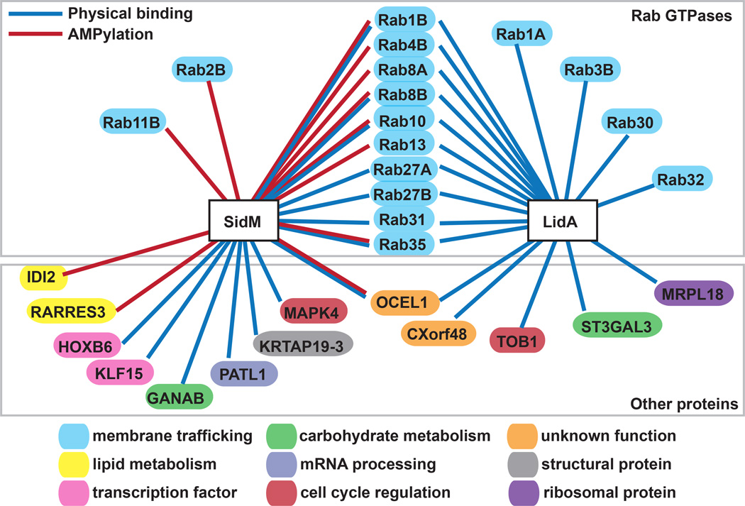
Host-pathogen protein interactions are fundamental to every microbial infection, yet their identification has remained challenging due to the lack of simple detection tools that avoid abundance biases while providing an open format for experimental modifications. Here, we applied the Nucleic Acid-Programmable Protein Array and a HaloTag-Halo ligand detection system to determine the interaction network of Legionella pneumophila effectors (SidM and LidA) with 10 000 unique human proteins. We identified known targets of these L. pneumophila proteins and potentially novel interaction candidates. In addition, we applied our Click chemistry-based NAPPA platform to identify the substrates for SidM, an effector with an adenylyl transferase domain that catalyzes AMPylation (adenylylation), the covalent addition of adenosine monophosphate (AMP). We confirmed a subset of the novel SidM and LidA targets in independent in vitro pull-down and in vivo cell-based assays, and provided further insight into how these effectors may discriminate between different host Rab GTPases. Our method circumvents the purification of thousands of human and pathogen proteins, and does not require antibodies against or prelabeling of query proteins. This system is amenable to high-throughput analysis of effectors from a wide variety of human pathogens that may bind to and/or post-translationally modify targets within the human proteome.

Keywords: AMPylation; LidA; Rab1; SidM; interactome; nucleic acid programmable protein array (NAPPA).


Figures

Figure 1
Figure 1
Optimization of the high-throughput NAPPA interaction assay for L. pneumophila effectors. (A) Flow scheme of Nucleic Acid Programmable Protein Array (NAPPA) fabrication and protein interaction assay. Plasmid cDNA of ~10,000 human genes was printed on aminosilane-coated slides at a density of ~2,000 genes per slide. DNA immobilization was validated with PicoGreen staining (green); display of recombinant tagged bait proteins was examined with appropriate anti-tag antibody (red) after IVTT. Binding of HaloTag-query protein to its interactor on NAPPA was detected using Alexa660-labeled Halo-ligand. (B) Comparison of protein interaction assay using the query protein LidA produced from E. coli-, wheat germ-, and human HeLa cell-based IVTT systems as well as N-terminal and C-terminal HaloTag constructs. Circles indicate the location of protein spots with enhanced signal. (C) Quantitative comparison of interactors for LidA sorted by their Z-score calculated from (B). (D) In-gel fluorescence analysis of in vitro translated LidA with N-terminal and C-terminal HaloTag.
Figure 2
Figure 2
Identification of host targets for L. pneumophila effectors by physical interaction in NAPPA. (A) Protein expression and display on NAPPA with (red) and without (green) T7 polymerase. (B) Correlation of protein expression and display on two identical NAPPA arrays. (C) Selection of host target candidates for L. pneumophila effectors using a Z-score threshold of 3. (D) Representative images of NAPPA slides probed with HaloTag (control) or HaloTag-LidA. Yellow boxes are shown enlarged in center panels, and regions of interest are marked with an arrow.
Figure 3
Figure 3
Confirmation of target candidates using in vitro bead-based pull-down assay. (A) Flow scheme of the high throughput pulldown assay. (B) Immunoblot analysis of the pulldown. Top panel (pulldown, PD) shows that the query proteins (HaloTag-LidA or -SidM), but not the Tag, were precipitated by GST-bait-coated beads. Bottom panel (western blot, WB) shows detection of the GST-bait protein by immunoblot using anti-GST antibody.
Figure 4
Figure 4
Conserved regions within Rab proteins targeted by LidA. Sequence alignment of Rab GTPases was performed using the MUSCLE server (http://www.bioinformatics.nl/tools/muscle.html). Rabs that interact with LidA are separated by a red line from those that do not interact. Regions of high homology are shown in magenta; regions of limited homology are shown in grey. Clusters of enhanced conservation are labeled by boxes. * indicates amino acid residues known to be involved in LidA-Rab1 binding (23).
Figure 5
Figure 5
Detection of AMPylated host targets by combining NAPPA and click chemistry. (A) Representative images of AMPylated targets (yellow boxes) on NAPPA slides after incubation with either wild-type SidM or its AMPylation-defective mutant SidMD110/112A, annotated as in Figure 2D. (B) Confirmation of target candidates using bead-based AMPylation assay with wild-type SidM (WT) or SidMD110/112A (D/A). Top panel (Az-rho) shows the detection of AMPylated targets using in-gel fluorescence with az-rho based on click reaction. Bottom panel (WB) is the detection of the host target protein (substrates) by immunoblot.
Figure 6
Figure 6
Host target validation using co-localization and co-precipitation analysis. (A) COS1 cells were transiently transfected with plasmids encoding mCherry-SidMD110/112A and GFP-tagged target candidates, and protein colocalization 16 hours after transfection was determined by fluorescence microscopy. Left panels show representative fluorographs of doubly transfected cells; the right panel shows line scans denoting pixel intensity of red and green fluorescent signals along the line indicated in the image to the left. Scale bar, 1µm. (B) Quantification of (A) showing the percentage of cells with coincident areas of GFP- and mCherry-enrichment. Data are mean ± SD (error bars) for three independent experiments. ****P < 0.0001 (two-tailed t-test). (C) Pulldown assay. Beads coated with BSA (control) or purified recombinant LidA were used to precipitate GFP-tagged prey proteins from 293T cell lysate. Inputs (1%) and eluates (50%) were separated by SDS-PAGE, and prey proteins were detected by immunoblot using anti-GFP antibody. Estimated molecular weights of prey proteins: GFP (27kD), GFP-Rab1B (52kD), GFP-Rab8B (51kD), GFP-Rab10 (50kD), GFP-Rab27A (52kD), GFP-OCEL1 (56kD). The graph below each panel is a quantification of the co-precipitation data. The amount of prey protein was determined by densitometry and is shown relative to nonspecifically-bound prey protein eluted by BSA-coated beads for each group, arbitrarily set at 1. The values are representative of at least two independent experiments.
Figure 7
Figure 7
Localization of host targets to the LCV. (A) CHO-FcγRII cells were transfected with constructs encoding the indicated GFP-tagged Rab proteins or OCEL1, and challenged with L. pneumophila Lp02 (wild-type) or Lp03 (T4SS mutant). Intracellular bacteria (red) were detected with anti-L. pneumophila antibody. Scale bar, 1µm. Line scans (right panels) denote pixel intensity of red and green fluorescent signals along the indicated line. (B) Quantification of (A) showing the percentage of cells displaying an LCV-specific GFP signal. Data are mean ± SD (error bars) for three independent experiments. **P < 0.01 (two-tailed t-test).
Figure 8
Figure 8
Host interaction profile for L. pneumophila SidM and LidA. Potential host targets for L. pneumophila SidM and LidA are mapped according to physical binding interaction (blue lines) or AMPylation (red lines). Targets are divided into Rab GTPase family members (top panel) or non-Rab GTPases (bottom panel), and color-coded according to protein function. Only those targets confirmed by bead-based NAPPA assay are shown.

References

    1. Ensminger AW, Isberg RR. Legionella pneumophila Dot/Icm translocated substrates: a sum of parts. Current opinion in microbiology. 2009;12(1):67–73. - PMC - PubMed
    1. Hubber A, Roy CR. Modulation of host cell function by Legionella pneumophila type IV effectors. Annual review of cell and developmental biology. 2010;26:261–283. - PubMed
    1. Vogel JP, Isberg RR. Cell biology of Legionella pneumophila. Curr Opin Microbiol. 1999;2(1):30–34. - PubMed
    1. Sherwood RK, Roy CR. A rab-centric perspective of bacterial pathogen-occupied vacuoles. Cell host & microbe. 2013;14(3):256–268. - PMC - PubMed
    1. Machner MP, Chen Y. Catch and release: Rab1 exploitation by Legionella pneumophila. Cell Logist. 2011;1(4):133–138. - PMC - PubMed

Publication types

MeSH terms

LinkOut - more resources