. 2015 Feb 12:13:56.
doi: 10.1186/s12967-015-0421-4.

Hypoxia-induced overexpression of stanniocalcin-1 is associated with the metastasis of early stage clear cell renal cell carcinoma

Affiliations

Hypoxia-induced overexpression of stanniocalcin-1 is associated with the metastasis of early stage clear cell renal cell carcinoma

Xin Ma et al. J Transl Med. .

Abstract

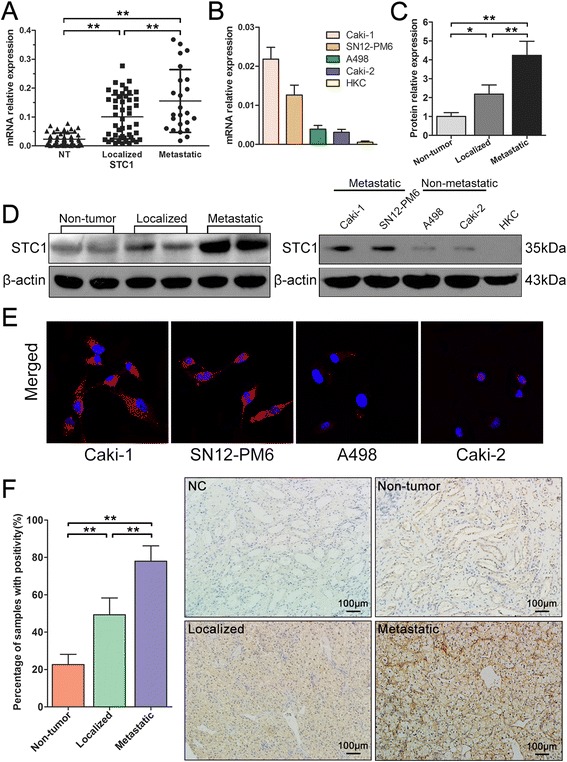
Background: Although metastasis of clear cell renal cell carcinoma (ccRCC) is predominantly observed in late stage tumors, early stage metastasis of ccRCC can also be found with indefinite molecular mechanism, leading to inappropriate clinical decisions and poor prognosis. Stanniocalcin-1 (STC1) is a glycoprotein hormone involved in calcium/phosphate homeostasis, which regulates various cellular processes in normal development and tumorigenesis. This study aimed to investigate the role and mechanism of regulation of STC1 in the metastasis of early stage ccRCC.

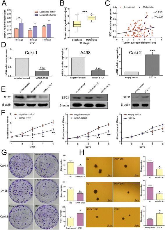
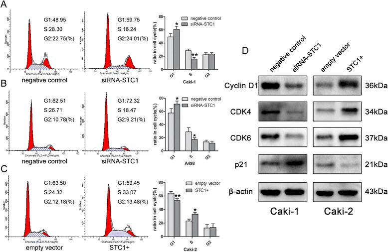
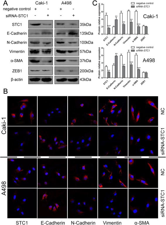
Methods: STC1 mRNA and protein expression was determined in ccRCC surgical specimens, RCC cell lines, and human kidney tubule epithelial cell line HKC by real-time polymerase chain reaction (RT-PCR) and western blotting. Immunohistochemistry staining (IHC) and immunofluorescence were also used to examine the expression and localization of STC1 in ccRCC tissues and cancer cells. Knockdown and overexpression studies were conducted in vitro in RCC cell lines using small interfering RNAs (siRNA) and lentiviral-mediated gene delivery to evaluate the role of STC1 in cell proliferation, anchorage-dependent and independent growth, cell cycle control, and migration and invasion.

Results: STC1 mRNA and protein expression were significantly up-regulated in tumors when compared with non-tumor tissues, with the greatest increase in expression observed in metastatic tissues. Clinicopathological analysis revealed that STC1 mRNA expression was associated with Fuhrman tumor grade (P = 0.008) and overall Tumor Node Metastasis (TNM) staging (P = 0.018). STC1 expression was elevated in T1 stage metastatic tumors when compared with localized tumors, and was positively correlated with average tumor diameter. Silencing of STC1 expression by Caki-1 and A498 resulted in the inhibition of cell proliferation, migration, and invasion, meanwhile down-regulation of STC1 impaired epithelial-mesenchymal transition (EMT) of ccRCC cell lines. Overexpression of STC1 in Caki-2 enhanced cell growth and proliferation but not migration and invasion. Further investigation identified hypoxia and HIF-1α as candidate regulators of STC1 expression.

Conclusions: Our findings demonstrate a role for STC1 in metastasis of early stage ccRCC and suggest that STC1 may be a biomarker of potential value both for the prognosis of this disease and for guiding clinical decisions regarding surgical strategies and adjuvant treatment.


Figures

Figure 1
Figure 1
Expression of stanniocalcin-1 (STC1) in clinical samples and renal cell carcinoma (RCC) cell lines. (A) The mRNA level of STC1 was significantly up-regulated in clear cell renal cell carcinoma (ccRCC) tissues compared with adjacent normal renal tissues, with the greatest expression observed in metastatic tissues. (B) The mRNA expression of STC1 was up-regulated in RCC cell lines compared with HKC-renal tubular epithelial cell line, with the greatest expression observed in metastatic cells. (C) (D) Western blotting showed the changes of protein expression were consistent with these of mRNA in clinical samples and RCC cell lines. (E) Immunofluorescence analysis revealed a significant increase in the intensity of STC1 staining in the cytoplasm of metastatic cell lines. (F) Representative images of STC1 immunohistochemistry staining in non-tumor (NT) tissues, localized and metastatic ccRCC tissues. Scale bar, 100 μm. Quantitative analysis of STC1 protein levels in the cytoplasm in 10 sets of paired NT tissues, localized and metastatic ccRCC tissues (n = 10). *,P < 0.05; **,P < 0.01.
Figure 2
Figure 2
Elevated expression of STC1 is associated with metastasis of T1 stage ccRCC. Effects of STC1 interferences on cell proliferation. (A) Analysis showing higher STC1 expression in T1 stage metastatic tumors when compared with localized tumors. (B) Analysis showing significantly larger average diameter in metastatic tumors (5.433 ± 0.644 cm, n = 10) when compared with localized tumors (3.833 ± 1.187 cm, n = 96). (C) Positive correlation of STC1 mRNA levels and average tumor diameter in T1 stage (n = 106, r = 0.215, p = 0.027). (D) (E) Alteration for STC1 mRNA and protein levels in Caki-1-Metastatic cell line, A498-Non-metastatic cell line and Caki-2-Non-metastatic cell line cells after knockdown or overexpression. (F) MTS assay showed STC1 knockdown reduced the proliferation of Caki-1 and A498 cells, while STC1 overexpression promoted the proliferation of Caki-2 cells. (G) (H) Effects of STC1 interferences on anchorage-dependent and independent growth of RCC cells. *,P < 0.05; **,P < 0.01; ***,P < 0.001.
Figure 3
Figure 3
Effect of STC1 interferences on cell cycle. (A) (B) Knockdown of STC1 led to G1 restoration compared with negative control group in Caki-1 and A498 cells. (C) Overexpression of STC1 induced G1-S phase transition compared to empty vector group in Caki-2 cells. (D) Western blotting results for the expression alteration of several key cell cycle regulatory factors. *,P < 0.05; **,P < 0.01.
Figure 4
Figure 4
Ability of STC1 in influencing the migration and invasion of ccRCC cells in vitro . (A) (B) Representative photographs of Transwell assays (magnification, ×100) in Caki-1 and A498 cells. (C) (D) Confluent monolayer cells were scratched using a sterile 200 μL pipette. Photos of the wound were taken at different time points (0, 12, and 24 h after scratching). STC1 knockdown impaired the cell mobility in Caki-1 and A498 cells. Each experiment was performed triplicates. *,P < 0.05; **,P < 0.01.
Figure 5
Figure 5
Knockdown of STC1 impairs epithelial-mesenchymal transition (EMT) of Caki-1 and A498 cells. (A) (B) Forty-eight hours after transfection, a series of EMT-related markers, including the mesenchymal markers N-cadherin, Vimentin, α-SMA, ZEB1, and the epithelial marker E-cadherin were determined by Western blotting and immunofluorescence staining (magnification, ×600) Red, CF555; blue, DAPI. (C) Western blotting results for the protein expression alteration of several EMT-related markers. **,P < 0.01; ***,P < 0.001.
Figure 6
Figure 6
Hypoxia induces STC1 expression. (A) The mRNA expression trend of STC1 and hypoxia inducible factor-1-alpha (HIF-1α) in ccRCC cell lines and HKC cells. (B) The association between STC1 and HIF-1α mRNA expression in clinical samples. (C) Representative images showed the morphological changes of RCC cell lines and HKC cells after CoCl2 treatment (magnification, ×100). (D) The mRNA level changes of STC1 and HIF-1α in A498 and Caki-2 cells after CoCl2 treatment. *,P < 0.05; **,P < 0.01; ***,P < 0.001.

Similar articles

Cited by

References

    1. Jemal A, Bray F, Center MM, Ferlay J, Ward E, Forman D. Global cancer statistics. CA Cancer J Clin. 2011;61:69–90. doi: 10.3322/caac.20107. - DOI - PubMed
    1. Kane CJ, Mallin K, Ritchey J, Cooperberg MR, Carroll PR. Renal cell cancer stage migration: analysis of the National Cancer Data Base. Cancer. 2008;113:78–83. doi: 10.1002/cncr.23518. - DOI - PubMed
    1. Ljungberg B, Cowan NC, Hanbury DC, Hora M, Kuczyk MA, Merseburger AS, et al. EAU guidelines on renal cell carcinoma: the 2010 update. Eur Urol. 2010;58:398–406. doi: 10.1016/j.eururo.2010.06.032. - DOI - PubMed
    1. Campbell SC, Novick AC, Belldegrun A, Blute ML, Chow GK, Derweesh IH, et al. Guideline for management of the clinical T1 renal mass. J Urol. 2009;182:1271–1279. doi: 10.1016/j.juro.2009.07.004. - DOI - PubMed
    1. Chang AC, Jellinek DA, Reddel RR. Mammalian stanniocalcins and cancer. Endocr Relat Cancer. 2003;10:359–373. doi: 10.1677/erc.0.0100359. - DOI - PubMed

Publication types

MeSH terms