Review
. 2016 May;91(2):452-68.
doi: 10.1111/brv.12178. Epub 2015 Mar 4.

De novo lipogenesis in the liver in health and disease: more than just a shunting yard for glucose

Affiliations
Review

De novo lipogenesis in the liver in health and disease: more than just a shunting yard for glucose

Francis W B Sanders et al. Biol Rev Camb Philos Soc. 2016 May.

Abstract

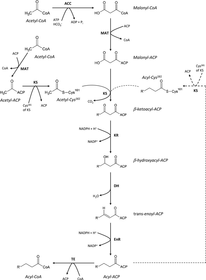
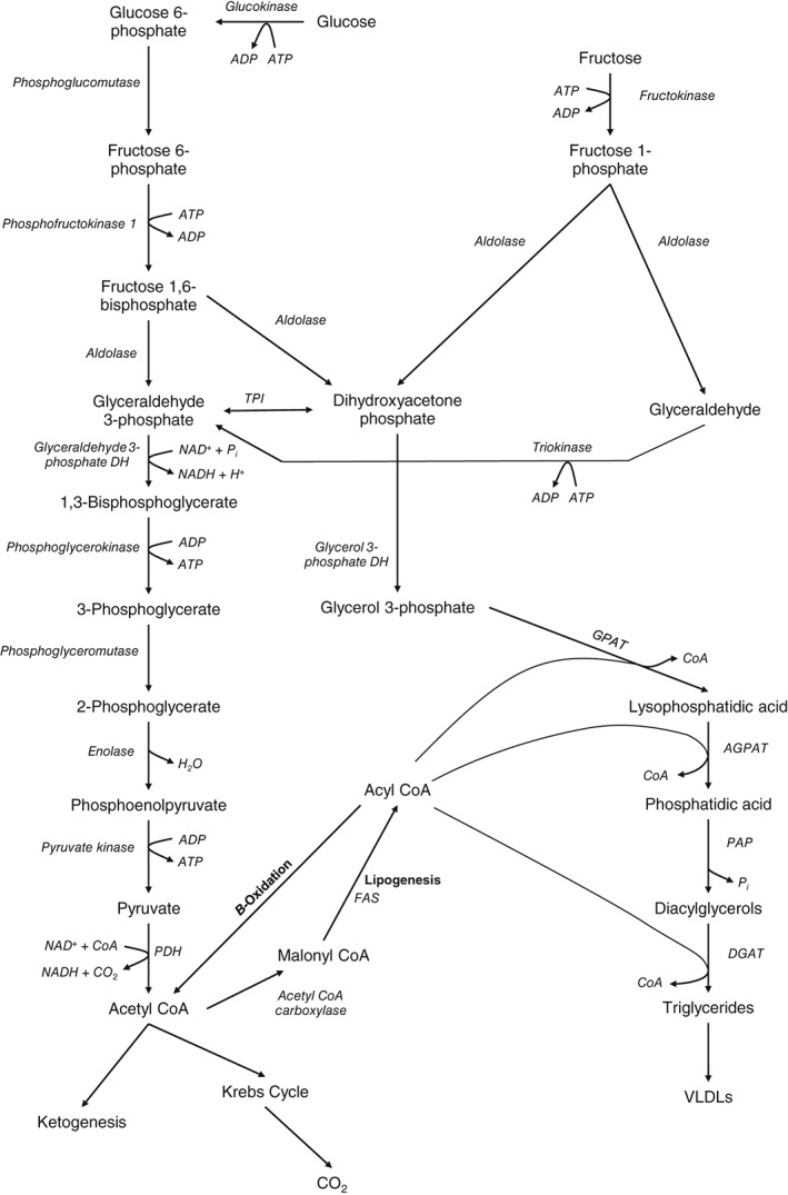
Hepatic de novo lipogenesis (DNL) is the biochemical process of synthesising fatty acids from acetyl-CoA subunits that are produced from a number of different pathways within the cell, most commonly carbohydrate catabolism. In addition to glucose which most commonly supplies carbon units for DNL, fructose is also a profoundly lipogenic substrate that can drive DNL, important when considering the increasing use of fructose in corn syrup as a sweetener. In the context of disease, DNL is thought to contribute to the pathogenesis of non-alcoholic fatty liver disease, a common condition often associated with the metabolic syndrome and consequent insulin resistance. Whether DNL plays a significant role in the pathogenesis of insulin resistance is yet to be fully elucidated, but it may be that the prevalent products of this synthetic process induce some aspect of hepatic insulin resistance.

Keywords: de novo lipogenesis (DNL); fructose; liver; non-alcoholic fatty liver disease (NAFLD); selective insulin resistance.


Figures

Figure 1
Figure 1
De novo lipogenesis. Malonyl‐CoA, as the additive monomer for DNL is generated from acetyl‐CoA under the catalytic activity of ACC. The acetyl and malonyl substrates for DNL are transferred to ACP. The acetyl‐CoA is then bound to the Cys161 of KS, followed by the decarboxylative condensation of acetyl‐CoA and malonyl‐CoA, forming β‐ketoacyl‐ACP catalysed by KS. The β‐ketone group of β‐ketoacyl‐ACP is reduced using the cosubstrate NADPH under the catalysis of KR, generating a β‐hydroxyacyl‐ACP intermediate. This reaction is followed by dehydration of the β‐carbon, producing a trans‐enoyl‐ACP intermediate. The trans double bond between the α‐ and β‐carbons is reduced under the catalytic action of EnR, using NAPDH as a substrate, forming acyl‐ACP. This can re‐enter the cycle to act as a substrate for KS, or when the R′ carbon chain is of sufficient length, the ACP is replaced with CoA and the acyl‐CoA released from FAS as TE is used to enzymatically cleave the acyl product from ACP. ACP, DH, EnR, KS, MAT and TE are all part of the multifunctional FAS. ACC, acetyl‐CoA carboxylase; ACP, acyl carrier protein; CoA, coenzyme A; DH, dehydratase; DNL, de novo lipogenesis; EnR, enoyl‐reductase; FAS, fatty acid synthase; KR, β‐ketoreductase; KS, β‐ketoacyl synthase; NADP+, nicotine adenine dinucleotide phosphate; NADPH, reduced form of NADP+; MAT, malonyl/acetyl transaferase; TE, thioesterase.
Figure 2
Figure 2
Hepatic glycolysis, fructolysis and triglyceride synthesis. Glucose undergoes the multistep process of glycolysis to generate pyruvate which is subsequently utilised for a range of cellular metabolic processes. This includes complete oxidative catabolism to carbon dioxide, lipogenesis under the catalysis of fatty acid synthase (FAS), and ketogenesis to generate ketone bodies for oxidative metabolic fuel. The fructolytic pathway feeds into the glycolytic pathway at the level of the trioses: dihydroxyacetone phosphate and glyceraldehyde 3‐phosphate. This thus bypasses the crucial regulatory steps in liver glycolytic metabolism, whereby glucokinase and phosphofructokinase‐1 are controlled by the metabolic milieu in coordination with hormonal effectors. This allows for the production of acetyl‐CoA from fructose to continue without regulation by insulin, and hence promote lipogenesis, and subsequent triglyceride (TG) synthesis from the acyl‐coenzyme A (CoA) product. This involves catalysis of progressive acylation of a glycerol‐phosphate backbone by glycerol‐phosphate acyl transferase (GPAT), acylglycerol‐phosphate acyl transferase (AGPAT). Dephosphorylation of the glycerol backbone then occurs under the catalysis of phosphatidic acid phosphorylase (PAP), followed by a further acylation of the diacylglycerol by diacylglycerol acyl transferase (DGAT). The TGs produced are packed into very low density lipoproteins (VLDLs) for export from the liver, or are stored within hepatocytes. DH, dehydrogenase; NAD+, nicotine adenine dinucleotide; NADH, reduced form of NAD+; PDH, pyruvate dehydrogenase; TPI, triose phosphate isomerase.
Figure 3
Figure 3
The liver acinus and zonation of metabolic processes. (A) The gross cytoarchitecture of the hepatic parenchyma. Hepatic lobules consist of roughly hexagonal acini with a central vein and portal triads at the interfaces of the acini. (B) A cross section of liver tissue along the portocentral axis, demonstrates the proposed zonation of metabolic processes, with the pericentral zone as the primary site of de novo lipogenesis (DNL) and the periportal zone as the primary site for gluconeogenesis. Endothelial cells make up the walls of the sinusoid which also contains macrophages known as Kupffer cells. As indicated by the arrows, blood flows from the portal area via the sinusoid into the hepatic venule. Bile flows in the opposite direction from hepatocytes to the bile duct through the bile canaliculi. Diagram based on Frevert et al. (2005).
Figure 4
Figure 4
Regulation of de novo lipogenesis by SREBP1c and ChREBP. Insulin activation of the insulin receptor leads to phosphorylation of insulin receptor substrate 1 (IRS1), which subsequently activates phosphoinositide 3‐kinase (PI3K), leading to the phosphorylation of phosphatidylinositol (4,5)‐bisphosphate (PIP2) to form phosphatidylinositol (3,4,5)‐trisphosphate (PIP3). PIP3 activates both phosphoinositide‐dependent kinase 1 (PDK1) and mammalian target of rapamycin complex 2 (mTORC2) (Gan et al., 2011). mTORC2 and PDK1 each lead to phosphorylation of protein kinase B (PKB), with mTORC2 promoting PKB‐mediated inhibition of glycogen synthase kinase 3β (GSK3β) (Hagiwara et al., 2012). PKB also activates mTORC1, leading to activation of ribosomal protein S6 kinase 1 (S6K1) which leads to nuclear localisation of liver X receptor α (LXRα) (Hwahng et al., 2009), heterodimerisation with retinoid X receptor (RXR) and subsequent transcription of lipogenic genes including sterol regulatory element binding protein 1c (SREBP1c). Full maturation of SREBP1c is promoted by the activity of mTORC1 through inhibition of lipin 1 (Peterson et al., 2011), as well as through inhibition of GSK3β by PKB. Both of these events leads to disinhibition of SREBP1c maturation and nuclear localisation. The nascent SREBP1c is a transmembrane protein in the lipid bilayer of the endoplamic reticulum (ER), associated with SREBP cleavage activated protein (SCAP), and insulin‐induced gene 1 (INSIG1). INSIG1 inhibits the SCAP‐mediated shuttling of SREBP1c to the Golgi apparatus via coat protein II (COPII)‐coated vesicles. When SREBP1c and SCAP are phosphorylated or sterols bind to SCAP it changes conformation dissociating from INSIG1 and allowing interaction with COPII coat proteins. SREBP1c is subsequently cleaved by two proteases, site 1‐protease (S1P) and site 2‐protease (S2P), in the Golgi leading to dissociation of SREBP1c from SCAP and removal of the transmembrane domain to allow the mature form of SREBP1c to localise to the nucleus. Once in the nucleus SREBP1c promotes transcription of lipogenic genes including fatty acid synthase (FAS), stearoyl‐CoA desaturase 1 (SCD1), elongation of long‐chain fatty acids family member 6 (ELOVL6), and acetyl CoA carboxylase (ACC). Glucose delivery to the hepatocyte also stimulated the transcription of lipogenic genes via the activation of carbohydrate response element binding protein (ChREBP). This occurs as glucose enters hepatocytes via glucose transporter 2 (GLUT2), and enters the glycolytic pathway. Initially glucose is phosphorylated to glucose 6‐phosphate (G6P), followed by isomerisation to fructose 6‐phosphate (F6P) and further phosphorylation to fructose‐2,6‐bisphosphate (F2,6P) through glycolysis. It has been suggested that both G6P and F2,6P through incompletely elucidated pathways lead to the dephosphorylation of ChREBP and dissociation from the cytosolic protein 14‐3‐3. This allows nuclear localisation of ChREBP and subsequent transcription of its target lipogenic genes, including FAS, SCD1, ELOVL6, ACC and pyruvate kinase, liver and RBC (Pklr).

References

    1. Aarsland, A. , Chinkes, D. & Wolfe, R. R. (1996). Contributions of de novo synthesis of fatty acids to total VLDL‐triglyceride secretion during prolonged hyperglycemia/hyperinsulinemia in normal man. Journal of Clinical Investigation 98, 2008–2017. - PMC - PubMed
    1. Abu‐Elheiga, L. , Brinkley, W. R. , Zhong, L. , Chirala, S. S. , Woldegiorgis, G. & Wakil, S. J. (2000). The subcellular localization of acetyl‐CoA carboxylase 2. Proceedings of the National Academy of Sciences of the United States of America 97, 1444–1449. - PMC - PubMed
    1. Abu‐Shanab, A. & Quigley, E. M. M. (2010). The role of the gut microbiota in nonalcoholic fatty liver disease. Nature Reviews Gastroenterology and Hepatology 7, 691–701. - PubMed
    1. Aguado, B. & Campbell, R. D. (1998). Characterization of a human lysophosphatidic acid acyltransferase that is encoded by a gene located in the class III region of the human major histocompatibility complex. Journal of Biological Chemistry 273, 4096–4105. - PubMed
    1. Ahmad, F. , Ahmad, P. M. , Pieretti, L. & Watters, G. T. (1978). Purification and subunit structure of rat mammary gland acetyl coenzyme A carboxylase. Journal of Biological Chemistry 253, 1733–1737. - PubMed

Publication types