. 2015 Mar 5:5:8769.
doi: 10.1038/srep08769.

Structure of human dipeptidyl peptidase 10 (DPPY): a modulator of neuronal Kv4 channels

Affiliations

Structure of human dipeptidyl peptidase 10 (DPPY): a modulator of neuronal Kv4 channels

Gustavo Arruda Bezerra et al. Sci Rep. .

Abstract

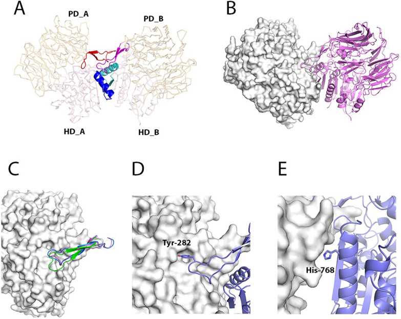
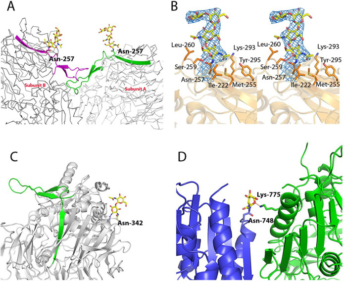
The voltage-gated potassium channel family (Kv) constitutes the most diverse class of ion channels in the nervous system. Dipeptidyl peptidase 10 (DPP10) is an inactive peptidase that modulates the electrophysiological properties, cell-surface expression and subcellular localization of voltage-gated potassium channels. As a consequence, DPP10 malfunctioning is associated with neurodegenerative conditions like Alzheimer and fronto-temporal dementia, making this protein an attractive drug target. In this work, we report the crystal structure of DPP10 and compare it to that of DPP6 and DPP4. DPP10 belongs to the S9B serine protease subfamily and contains two domains with two distinct folds: a β-propeller and a classical α/β-hydrolase fold. The catalytic serine, however, is replaced by a glycine, rendering the protein enzymatically inactive. Difference in the entrance channels to the active sites between DPP10 and DPP4 provide an additional rationale for the lack of activity. We also characterize the DPP10 dimer interface focusing on the alternative approach for designing drugs able to target protein-protein interactions.


Figures

Figure 1
Figure 1. Overall structure of DPP10.
(A) Scheme displaying the domain organization of DPP10. The domains are represented by boxes: the cytoplasmic domain is shown in cyan, the transmembrane domain in green, the β-propeller in yellow, the α/β-hydrolase domain in red and the extended arm in blue. (B) Cartoon representation of the structure of DPP10 using the same color code as in panel A. Sugar residues are shown as yellow sticks. (C) Comparison of the structures of DPP10 (light red), DPP6 (blue) and DPP4 (yellow) each in a cartoon representation.
Figure 2
Figure 2. “Catalytic triad” of DPP10.
(A) Superposition of DPP10 (shown in green) “catalytic triad” (Gly-651, His-759 and Asp-727), N-anchor glutamates (Glu-233 and Glu-234) and oxyanion holes (Asp-568 and Tyr 652) to that of DPP4 (shown in blue, PDB-code: 3EIO). (B) DPP10 “active site” 2Fo-Fc map contoured at 1σ is shown in blue. Side chains of the residues are shown as sticks.
Figure 3
Figure 3. Comparison of the “active site” access channel in DPP4, DPP10 and DPP6.
(A) Surface representation of DPP6, DPP10 and DPP4 with the channels shown in green, red and blue, respectively. The β-propeller domain is colored in orange and the α/β-hydrolase domain in pink. (B) Channel profiles indicating the radius in Å vs. the scaled length, starting from the “catalytic triad” position (indicated by a star) towards the protein surface (indicated by an arrow). Surface representation of the channels using the same coloring scheme as in panel A.
Figure 4
Figure 4. Dimerization interface.
(A) Cartoon representation of residues participating in the dimer interface. The β-propeller domains are indicated as PD_A and PD_B, and the α/β-hydrolase domains as HD_A and HD_B in subunits A and B, respectively. The β-propeller loops are colored in red and magenta, and the C-terminal helices in blue and cyan for the subunits A and B, respectively. (B) Heterodimer model of DPP10 (surface representation) and DPP6 (cartoon representation). This model was generated by superimposing DPP6 chain A onto DPP10 chain B. (C) Cartoon representation of β-propeller loop of DPP10 (blue), DPP6 (magenta) and DPPIV (green) superimposed to the opposite subunit of DPP10 (surface representation). (D) Interaction of Tyr-282 with the opposite subunit A in DPP10 and (E) His-768.
Figure 5
Figure 5. DPP10 glycosylation.
(A) The glycosylated Asn-257 is placed adjacent to the β-propeller loop, and might stabilize it. Both subunits are represented as ribbons, the β-propeller loops as cartoons and Asn-257 is shown as sticks. Subunit A is shown in light grey and green, while subunit B is shown in dark grey and magenta. (B) Interactions between the carbohydrate and the residues in the subunit A. Ser-229 makes a hydrogen bond with O5 (possibly O6 in subunit B) while Leu-222, Met-255, Leu-260, Tyr-295 and Lys-293 contribute with hydrophobic interactions. Glycan 2Fo-Fc map contoured at 1σ is shown in blue. (C) The glycosylation at Asn-342 might indirectly stabilize the β-propeller loop, since it is positioned relatively close to this region. The protein is shown as light grey cartoon and the β-propeller loop is shown as green cartoon. (D) N-acetylglucosamine bound to Asn-748 in the subunit B (shown as dark grey cartoon) possibly interacts through a hydrogen bond with Lys-775 of subunit A (shown as light grey cartoon). The carbohydrates are shown as yellow sticks and the hydrogen bond as red dashes.

References

    1. Ren X., Hayashi Y., Yoshimura N. & Takimoto K. Transmembrane interaction mediates complex formation between peptidase homologues and Kv4 channels. Mol Cell Neurosci 29, 320–32 (2005). - PubMed
    1. Herson P. S. & Adelman J. P. It takes two to tango, but three to ISA. Neuron 37, 370–2 (2003). - PubMed
    1. Covarrubias M. et al. The neuronal Kv4 channel complex. Neurochem Res 33, 1558–67 (2008). - PMC - PubMed
    1. Amberg G. C., Koh S. D., Imaizumi Y., Ohya S. & Sanders K. M. A-type potassium currents in smooth muscle. Am J Physiol Cell Physiol 284, C583–95 (2003). - PubMed
    1. Lundby A. et al. Effect of the I(to) activator NS5806 on cloned K(V)4 channels depends on the accessory protein KChIP2. Br J Pharmacol 160, 2028–44. - PMC - PubMed

Publication types

MeSH terms

Substances

Associated data