Acute ER stress regulates amyloid precursor protein processing through ubiquitin-dependent degradation
- PMID: 25740315
- PMCID: PMC5390087
- DOI: 10.1038/srep08805
Acute ER stress regulates amyloid precursor protein processing through ubiquitin-dependent degradation
Abstract
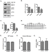
Beta-amyloid (Aβ), a major pathological hallmark of Alzheimer's disease (AD), is derived from amyloid precursor protein (APP) through sequential cleavage by β-secretase and γ-secretase enzymes. APP is an integral membrane protein, and plays a key role in the pathogenesis of AD; however, the biological function of APP is still unclear. The present study shows that APP is rapidly degraded by the ubiquitin-proteasome system (UPS) in the CHO cell line in response to endoplasmic reticulum (ER) stress, such as calcium ionophore, A23187, induced calcium influx. Increased levels of intracellular calcium by A23187 induces polyubiquitination of APP, causing its degradation. A23187-induced reduction of APP is prevented by the proteasome inhibitor MG132. Furthermore, an increase in levels of the endoplasmic reticulum-associated degradation (ERAD) marker, E3 ubiquitin ligase HRD1, proteasome activity, and decreased levels of the deubiquitinating enzyme USP25 were observed during ER stress. In addition, we found that APP interacts with USP25. These findings suggest that acute ER stress induces degradation of full-length APP via the ubiquitin-proteasome proteolytic pathway.
Conflict of interest statement
The authors declare no competing financial interests.
Figures


Similar articles
-
[Physiological Roles of Ubiquitin Ligases Related to the Endoplasmic Reticulum].Yakugaku Zasshi. 2016;136(6):805-9. doi: 10.1248/yakushi.15-00292-2. Yakugaku Zasshi. 2016. PMID: 27252059 Review. Japanese.
-
Loss of HRD1-mediated protein degradation causes amyloid precursor protein accumulation and amyloid-beta generation.J Neurosci. 2010 Mar 17;30(11):3924-32. doi: 10.1523/JNEUROSCI.2422-09.2010. J Neurosci. 2010. PMID: 20237263 Free PMC article.
-
CHIP stabilizes amyloid precursor protein via proteasomal degradation and p53-mediated trans-repression of β-secretase.Aging Cell. 2015 Aug;14(4):595-604. doi: 10.1111/acel.12335. Epub 2015 Mar 13. Aging Cell. 2015. PMID: 25773675 Free PMC article.
-
Cereblon-mediated degradation of the amyloid precursor protein via the ubiquitin-proteasome pathway.Biochem Biophys Res Commun. 2020 Mar 26;524(1):236-241. doi: 10.1016/j.bbrc.2020.01.078. Epub 2020 Jan 23. Biochem Biophys Res Commun. 2020. PMID: 31983437
-
[Molecular pharmacological studies on the protection mechanism against endoplasmic reticulum stress-induced neurodegenerative disease].Yakugaku Zasshi. 2012;132(12):1437-42. doi: 10.1248/yakushi.12-00249. Yakugaku Zasshi. 2012. PMID: 23208051 Review. Japanese.
Cited by
-
Aβ42 oligomers modulate β-secretase through an XBP-1s-dependent pathway involving HRD1.Sci Rep. 2016 Nov 17;6:37436. doi: 10.1038/srep37436. Sci Rep. 2016. PMID: 27853315 Free PMC article.
-
A Pipeline for Natural Small Molecule Inhibitors of Endoplasmic Reticulum Stress.Front Pharmacol. 2022 Jul 22;13:956154. doi: 10.3389/fphar.2022.956154. eCollection 2022. Front Pharmacol. 2022. PMID: 35935873 Free PMC article.
-
Advances in the study of protein folding and endoplasmic reticulum-associated degradation in mammal cells.J Zhejiang Univ Sci B. 2024 Mar 15;25(3):212-232. doi: 10.1631/jzus.B2300403. J Zhejiang Univ Sci B. 2024. PMID: 38453636 Free PMC article. Review.
-
Emerging Roles of Ubiquitin-Specific Protease 25 in Diseases.Front Cell Dev Biol. 2021 Jun 23;9:698751. doi: 10.3389/fcell.2021.698751. eCollection 2021. Front Cell Dev Biol. 2021. PMID: 34249948 Free PMC article. Review.
-
Usp25m protease regulates ubiquitin-like processing of TUG proteins to control GLUT4 glucose transporter translocation in adipocytes.J Biol Chem. 2018 Jul 6;293(27):10466-10486. doi: 10.1074/jbc.RA118.003021. Epub 2018 May 17. J Biol Chem. 2018. PMID: 29773651 Free PMC article.
References
-
- Murray F. E. et al. Elemental analysis of neurofibrillary tangles in Alzheimer's disease using proton-induced X-ray analysis. Ciba Found Symp 169, 201–210; discussion 210–206 (1992). - PubMed
-
- Sennvik K. et al. Calcium ionophore A23187 specifically decreases the secretion of beta-secretase cleaved amyloid precursor protein during apoptosis in primary rat cortical cultures. J Neurosci Res 63, 429–437 (2001). - PubMed
-
- Querfurth H. W. & Selkoe D. J. Calcium ionophore increases amyloid beta peptide production by cultured cells. Biochemistry 33, 4550–4561 (1994). - PubMed
Publication types
MeSH terms
Substances
LinkOut - more resources
Full Text Sources
Other Literature Sources

