Review
. 2015 Apr 7;282(1804):20142957.
doi: 10.1098/rspb.2014.2957.

An out-of-body experience: the extracellular dimension for the transmission of mutualistic bacteria in insects

Affiliations
Review

An out-of-body experience: the extracellular dimension for the transmission of mutualistic bacteria in insects

Hassan Salem et al. Proc Biol Sci. .

Abstract

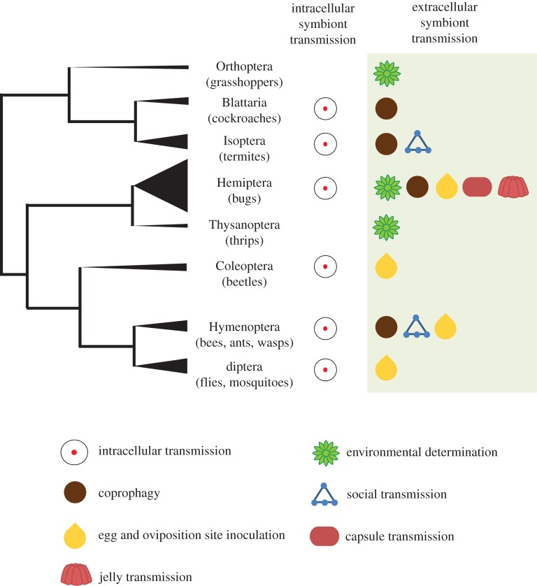
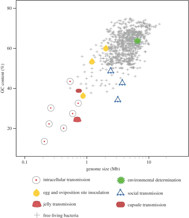
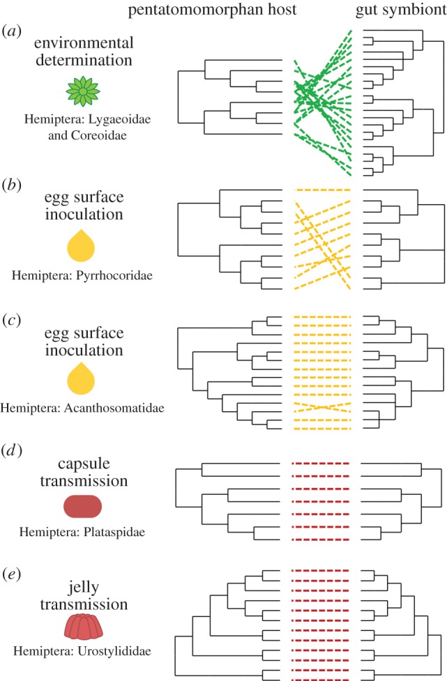
Across animals and plants, numerous metabolic and defensive adaptations are a direct consequence of symbiotic associations with beneficial microbes. Explaining how these partnerships are maintained through evolutionary time remains one of the central challenges within the field of symbiosis research. While genome erosion and co-cladogenesis with the host are well-established features of symbionts exhibiting intracellular localization and transmission, the ecological and evolutionary consequences of an extracellular lifestyle have received little attention, despite a demonstrated prevalence and functional importance across many host taxa. Using insect-bacteria symbioses as a model, we highlight the diverse routes of extracellular symbiont transfer. Extracellular transmission routes are unified by the common ability of the bacterial partners to survive outside their hosts, thereby imposing different genomic, metabolic and morphological constraints than would be expected from a strictly intracellular lifestyle. We emphasize that the evolutionary implications of symbiont transmission routes (intracellular versus extracellular) do not necessarily correspond to those of the transmission mode (vertical versus horizontal), a distinction of vital significance when addressing the genomic and physiological consequences for both host and symbiont.

Keywords: host–microbe coevolution; mutualism stability; symbiont transmission; symbiosis.


Figures

Figure 1.
Figure 1.
Cladogram depicting the diversity of insect orders with reported extracellularly transmitted bacterial symbionts (as listed in the electronic supplementary material, table S1). Symbols indicate extracellular transmission routes. Terminal branch thickness is proportional to the number of families within the order that have been reported to rely on an extracellular route for symbiont transfer. Orders featuring taxa with an intracellular symbiont transmission route are designated with a symbol as well (per electronic supplementary material, table S3). (Online version in colour.)
Figure 2.
Figure 2.
Extracellular symbiont transmission routes in insects. Horizontal transmission of Rickettsia among whiteflies (Bemisia tabaci) (a) involves the use of the insect's host plant [13] (b). Transmission of beneficial gut symbionts in the European firebug (c) relies on secretions that are smeared over the egg surface following oviposition (d,e) [14]. Beewolves (f) cultivate the defensive symbiont ‘Candidatus Streptomyces philanthi’ in specialized antennal gland reservoirs (g,h) and transmit it via the brood cell [15]. Fungus-growing ants harbour defensive bacteria in specialized regions of their cuticle (i,j) that are transmitted via social behaviour among nest-mates [16]. Beneath their egg mass, plataspid stinkbugs (k) deposit brown symbiont-bearing capsules (l) that are ingested by newly hatched nymphs (m) to initiate infection with the gut symbiont. An adult female of Urostylis westwoodii depositing egg-encapsulating, symbiont-containing jelly (n) that is later ingested by newly hatched nymphs (o). (Online version in colour.)
Figure 3.
Figure 3.
Relationship between genome size and GC content for a representative subset of intra- as well as all extracellularly transmitted bacterial symbionts in insects (per electronic supplementary material, table S2), as compared to free-living bacteria. Symbols indicate symbiont transmission route and biotic condition (symbiotic versus free-living). (Online version in colour.)
Figure 4.
Figure 4.
Comparison of evolutionary relationships between bugs and their gut symbionts as it relates to the symbionts' extracellular transmission routes. These relationships were established for (a) Lygaeoidea and Coreoidea and their environmentally acquired Burkholderia symbionts [20], (b) Pyrrhocoridae [64] and (c) Acanthosomatidae [39] relying on egg smearing, as well as (d) Plataspidae [43] and (e) Urostylididae [44] using symbiont capsules and jelly, respectively, for the transmission of their gut symbionts. (Online version in colour.)

Similar articles

Cited by

References

    1. McFall-Ngai M, et al. 2013. Animals in a bacterial world, a new imperative for the life sciences. Proc. Natl Acad. Sci. USA 110, 3229–3236. (10.1073/pnas.1218525110) - DOI - PMC - PubMed
    1. Buchner P. 1965. Endosymbiosis of animals with plant microorganisms. New York, NY: Interscience.
    1. Ebert D. 2013. The epidemiology and evolution of symbionts with mixed-mode transmission. Annu. Rev. Ecol. Evol. Syst. 44, 623–643. (10.1146/annurev-ecolsys-032513-100555) - DOI
    1. Baumann P. 2005. Biology of bacteriocyte-associated endosymbionts of plant sap-sucking insects. Annu. Rev. Microbiol. 59, 155–189. (10.1146/annurev.micro.59.030804.121041) - DOI - PubMed
    1. Moran NA, McCutcheon JP, Nakabachi A. 2008. Genomics and evolution of heritable bacterial symbionts. Ann. Rev. Genet. 42, 165–190. (10.1146/annurev.genet.41.110306.130119) - DOI - PubMed

Publication types