. 2015 Jul;96(Pt 7):1702-11.
doi: 10.1099/vir.0.000113. Epub 2015 Mar 4.

Physico-chemical requirements and kinetics of membrane fusion of flavivirus-like particles

Affiliations

Physico-chemical requirements and kinetics of membrane fusion of flavivirus-like particles

Danillo L A Espósito et al. J Gen Virol. 2015 Jul.

Abstract

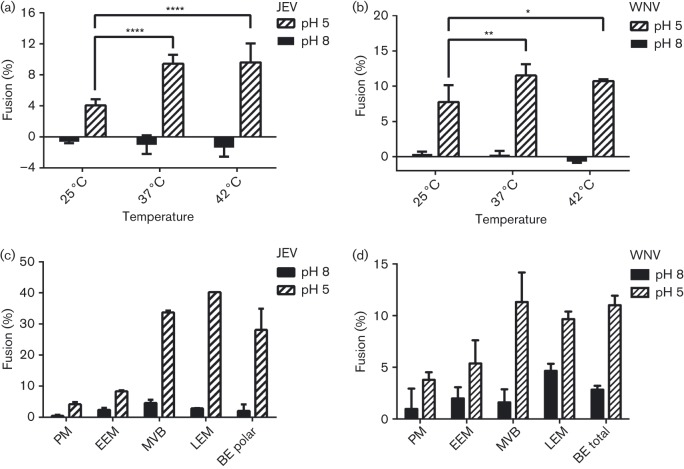
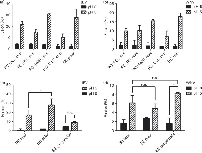
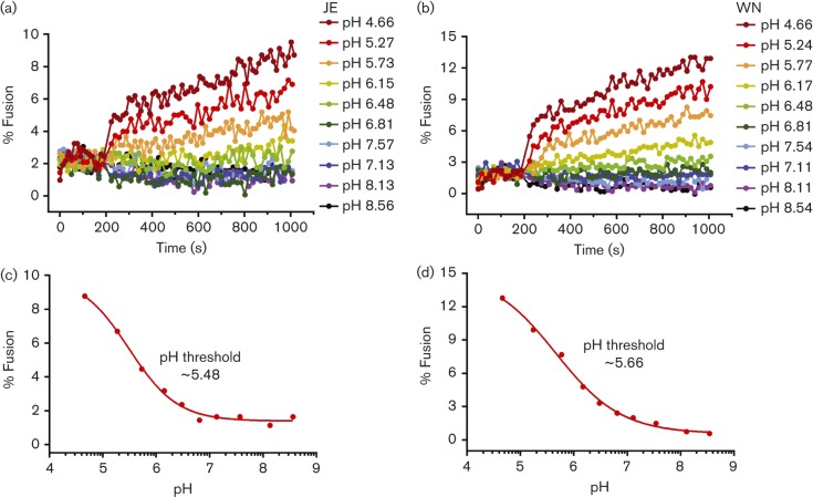
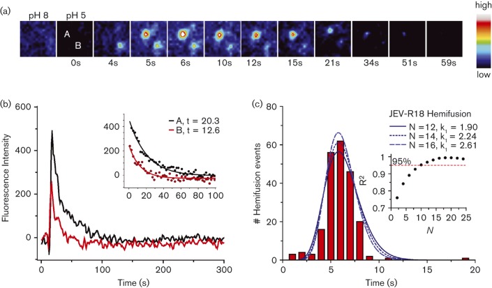
Flaviviruses deliver their RNA genome into the host-cell cytoplasm by fusing their lipid envelope with a cellular membrane. Expression of the flavivirus pre-membrane and envelope glycoprotein genes in the absence of other viral genes results in the spontaneous assembly and secretion of virus-like particles (VLPs) with membrane fusion activity. Here, we examined the physico-chemical requirements for membrane fusion of VLPs from West Nile and Japanese encephalitis viruses. In a bulk fusion assay, optimal hemifusion (or lipid mixing) efficiencies were observed at 37 °C. Fusion efficiency increased with decreasing pH; half-maximal hemifusion was attained at pH 5.6. The anionic lipids bis(monoacylglycero)phosphate and phosphatidylinositol-3-phosphate, when present in the target membrane, significantly enhanced fusion efficiency, consistent with the emerging model that flaviviruses fuse with intermediate-to-late endosomal compartments, where these lipids are most abundant. In a single-particle fusion assay, VLPs catalysed membrane hemifusion, tracked as lipid mixing with the cellular membrane, on a timescale of 7-20 s after acidification. Lipid mixing kinetics suggest that hemifusion is a kinetically complex, multistep process.


Figures

Fig. 1.
Fig. 1.. Membrane fusion efficiency of JEV and WNV VLPs as a function of temperature and lipid composition. (a, b) Fusion efficiency, measured as lipid mixing, of JEV (a) and WNV (b) VLPs is greater at 37 and 42 °C than at 25 °C. (c, d) Fusion efficiency of JEV (c) and WNV (d) VLPs with liposomes with compositions mimicking different cellular compartments. PM, plasma membrane; EEM, early endosomal membranes; MVB, multivesicular bodies; LEM, late endosomal membranes; BE, porcine brain extract lipids. Each value is the mean±standard deviation of triplicate measurements.
Fig. 2.
Fig. 2.. Role of polar and anionic lipids in VLP fusion. (a, b) Fusion efficiency of JEV (a) and WNV (b) VLPs with liposomes containing a ternary mixture of phosphatidylcholine (PC), with BMP, phosphatidylglycerol (PG), phosphatidylserine (PS), ceramide-1-phosphate (C1P) or ceramide (Cer), together and cholesterol (chol) at a 4 : 4 : 2 molar ratio. (c, d) Fusion efficiency of JEV (c) and WNV (d) VLPs with liposomes composed of total porcine brain extract lipids (BE total), polar brain lipids (BE polar) or ganglioside brain lipids (BE ganglioside).
Fig. 3.
Fig. 3.. Membrane fusion efficiency of JEV and WNV VLPs as a function of pH. (a, b) Fusion extent as a function of time post-acidification for JEV (a) VLPs and WNV (b) VLPs in SPG buffer (see Methods) at the indicated pH values. Curves were normalized to the fluorescence obtained after detergent solubilization of the self-quenched DiI in the VLPs. (c, d) End-point representations of the curves in (a) and (b), respectively, plotting fusion extent as a function of pH. Half-maximal fusion was observed at ~pH 5.5 for JEV VLPs and ~pH 5.7 for WNV VLPs.
Fig. 4.
Fig. 4.. TIRFM of membrane hemifusion by single JEV VLPs. (a) Snapshots (after bilinear interpolation) of two R18-labelled JEV VLPs fusing with the planar membrane. Images were false coloured with the indicated pixel value map. (b) Fluorescence intensities of the particles in (a) over the time course of fusion. Lipid mixing caused sudden dequenching of R18, followed by decay due to diffusion of the dye into the membrane. The exponential decay constant was 13–20 s (inset). (c) Distribution of time elapsed between pH drop and hemifusion of individual particles (n = 214). The best fit to a gamma distribution function was with N = 15 transitions, k 1 = 2.43 s−1. Inset, R2 values of gamma distribution fits at various N values.

References

    1. Acosta E. G., Castilla V., Damonte E. B. ( 2009. ). Alternative infectious entry pathways for dengue virus serotypes into mammalian cells. Cell Microbiol 11, 1533–1549. 10.1111/j.1462-5822.2009.01345.x - DOI - PMC - PubMed
    1. Bajjalieh S. M., Martin T. F., Floor E. ( 1989. ). Synaptic vesicle ceramide kinase. A calcium-stimulated lipid kinase that co-purifies with brain synaptic vesicles. J Biol Chem 264, 14354–14360. - PubMed
    1. Chao L. H., Klein D. E., Schmidt A. G., Peña J. M., Harrison S. C. ( 2014. ). Sequential conformational rearrangements in flavivirus membrane fusion. Elife 3, e04389. 10.7554/eLife.04389 - DOI - PMC - PubMed
    1. Chen Y., Maguire T., Hileman R. E., Fromm J. R., Esko J. D., Linhardt R. J., Marks R. M. ( 1997. ). Dengue virus infectivity depends on envelope protein binding to target cell heparan sulfate. Nat Med 3, 866–871. 10.1038/nm0897-866 - DOI - PubMed
    1. Das S., Chakraborty S., Basu A. ( 2010. ). Critical role of lipid rafts in virus entry and activation of phosphoinositide 3′ kinase/Akt signaling during early stages of Japanese encephalitis virus infection in neural stem/progenitor cells. J Neurochem 115, 537–549. 10.1111/j.1471-4159.2010.06951.x - DOI - PubMed

Publication types

LinkOut - more resources