. 2015 May;89(10):5569-80.
doi: 10.1128/JVI.00309-15. Epub 2015 Mar 4.

Characterization of the Influence of Semen-Derived Enhancer of Virus Infection on the Interaction of HIV-1 with Female Reproductive Tract Tissues

Affiliations

Characterization of the Influence of Semen-Derived Enhancer of Virus Infection on the Interaction of HIV-1 with Female Reproductive Tract Tissues

Shannon A Allen et al. J Virol. 2015 May.

Abstract

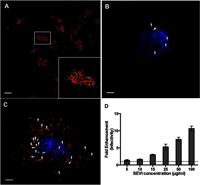
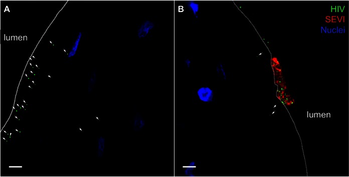
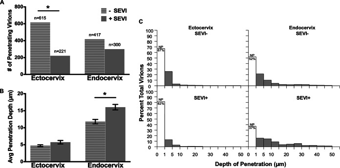
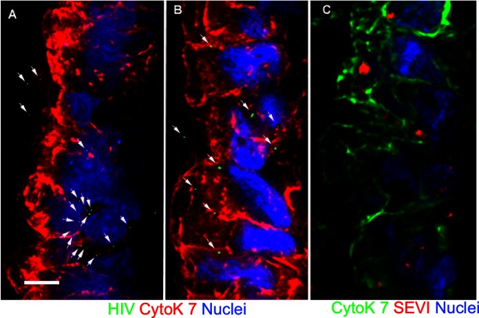
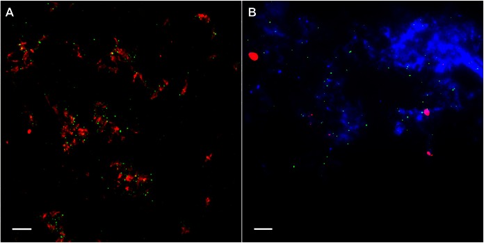
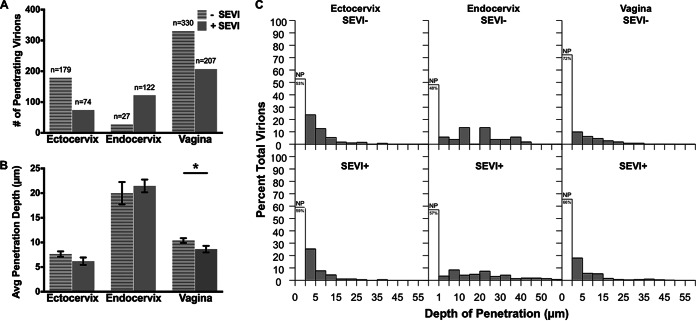
The majority of human immunodeficiency virus type 1 (HIV-1) transmission events occur in women when semen harboring infectious virus is deposited onto the mucosal barriers of the vaginal, ectocervical, and endocervical epithelia. Seminal factors such as semen-derived enhancer of virus infection (SEVI) fibrils were previously shown to greatly enhance the infectivity of HIV-1 in cell culture systems. However, when SEVI is intravaginally applied to living animals, there is no effect on vaginal transmission. To define how SEVI might function in the context of sexual transmission, we applied HIV-1 and SEVI to intact human and rhesus macaque reproductive tract tissues to determine how it influences virus interactions with these barriers. We show that SEVI binds HIV-1 and sequesters most virions to the luminal surface of the stratified squamous epithelium, significantly reducing the number of virions that penetrated the tissue. In the simple columnar epithelium, SEVI was no longer fibrillar in structure and was detached from virions but allowed significantly deeper epithelial virus penetration. These observations reveal that the action of SEVI in intact tissues is very different in the anatomical context of sexual transmission and begin to explain the lack of stimulation of infection observed in the highly relevant mucosal transmission model.

Importance: The most common mode of HIV-1 transmission in women occurs via genital exposure to the semen of HIV-infected men. A productive infection requires the virus to penetrate female reproductive tract epithelial barriers to infect underlying target cells. Certain factors identified within semen, termed semen-derived enhancers of virus infection (SEVI), have been shown to significantly enhance HIV-1 infectivity in cell culture. However, when applied to the genital tracts of living female macaques, SEVI did not enhance virus transmission. Here we show that SEVI functions very differently in the context of intact mucosal tissues. SEVI decreases HIV-1 penetration of squamous epithelial barriers in humans and macaques. At the mucus-coated columnar epithelial barrier, the HIV-1/SEVI interaction is disrupted. These observations suggest that SEVI may not play a significant stimulatory role in the efficiency of male-to-female sexual transmission of HIV.


Figures

FIG 1
FIG 1
SEVI fibrils bind HIV-1 and enhance infection in vitro. (A) SEVI fibrils (red) were incubated with HIV-1 (green). (B and C) TZM-bl reporter cells, visualized by Hoechst staining (blue), were incubated with either HIV-1 alone (B) or HIV-1/SEVI complexes obtained from the assay depicted in panel A (C). (D) Effect of SEVI on HIV-1 infectivity. Data were normalized by control conditions (cells only, AZT only, and SEVI only). The dotted line represents the 0-μg/ml SEVI conditions. Fold enhancement of infectivity was obtained from 3 separate experiments performed in triplicate. Arrows in panels B and C highlight HIV-1 on cells. Images were taken at a ×100 magnification. Bars = 5 μm.
FIG 2
FIG 2
SEVI sequesters HIV-1 to the luminal side of human ectocervical epithelium. Labeled SEVI fibrils were incubated with PA-HIV. Either PA-HIV alone (A) or PA-HIV-1/SEVI complexes (B) were incubated with ectocervical explants. Arrows point to areas of HIV-1 ingress into the epithelium. Epithelial nuclei are identified by Hoechst staining. All images were taken at a ×100 magnification. Bars = 5 μm.
FIG 3
FIG 3
SEVI differentially influences the numbers and depths of penetrating virions in human cervical tissues. (A and B) Bar graphs of the number of penetrating virions (A) and average virus penetration (B) with and without SEVI treatment for ectocervix and endocervix. (C) Histograms of the distributions of penetration depths for untreated (top) and SEVI-treated (bottom) HIV-1 in the ectocervix (left) and endocervix (right). The unshaded bars within each histogram show the percentages of nonpenetrators (NP), i.e., virions that penetrated to depths of <1 μm.
FIG 4
FIG 4
Untreated and SEVI-treated HIV-1 particles enter the epithelium similarly in human endocervical explants. SEVI fibrils were preincubated with PA–HIV-1. (A and B) Either PA–HIV-1 alone (A) or PA–HIV-1/SEVI complexes (B) were incubated with endocervical explants. (C) Physical appearance of SEVI within the simple columnar epithelium of the endocervix. Arrows in panels A and B highlight HIV-1 within the epithelium. The simple columnar epithelium is visualized by cytokeratin 7 staining and nuclear Hoechst staining. For each panel, the lumen is on the left side of the image. Images were taken at a ×100 magnification. Bars = 5 μm.
FIG 5
FIG 5
HIV-1 detaches from SEVI fibrils after inoculation with endocervical tissues. Shown are images of PA-HIV (green) and SEVI (red) complexes on a coverslip placed underneath ectocervical explants (A) and endocervical explants actively secreting mucus (blue) (B) during culture. Representative images from each tissue type (ectocervix and endocervix) reflect similar findings for all 12 donors. Images were taken at a ×100 magnification. Bars = 5 μm.
FIG 6
FIG 6
Virus penetration is dependent on tissue type and not SEVI in macaque genital tissues. (A and B) Bar graphs of the number of penetrating virions (A) and average virus penetration (B) with and without SEVI treatment for macaque ectocervix, endocervix, and vagina. (C) Histograms of penetration depths for untreated (top) and SEVI-treated (bottom) HIV-1 in macaque genital tissues (ectocervix [left], endocervix [middle], and vagina [right]). The unshaded bars in each panel show the percentages of nonpenetrators (NP).

References

    1. Kim KA, Yolamanova M, Zirafi O, Roan NR, Staendker L, Forssmann WG, Burgener A, Dejucq-Rainsford N, Hahn BH, Shaw GM, Greene WC, Kirchhoff F, Munch J. 2010. Semen-mediated enhancement of HIV infection is donor-dependent and correlates with the levels of SEVI. Retrovirology 7:55. doi:10.1186/1742-4690-7-55. - DOI - PMC - PubMed
    1. Munch J, Rucker E, Standker L, Adermann K, Goffinet C, Schindler M, Wildum S, Chinnadurai R, Rajan D, Specht A, Gimenez-Gallego G, Sanchez PC, Fowler DM, Koulov A, Kelly JW, Mothes W, Grivel JC, Margolis L, Keppler OT, Forssmann WG, Kirchhoff F. 2007. Semen-derived amyloid fibrils drastically enhance HIV infection. Cell 131:1059–1071. doi:10.1016/j.cell.2007.10.014. - DOI - PubMed
    1. Bouvet JP, Gresenguet G, Belec L. 1997. Vaginal pH neutralization by semen as a cofactor of HIV transmission. Clin Microbiol Infect 3:19–23. doi:10.1111/j.1469-0691.1997.tb00246.x. - DOI - PubMed
    1. Tevi-Benissan C, Belec L, Levy M, Schneider-Fauveau V, Si Mohamed A, Hallouin MC, Matta M, Gresenguet G. 1997. In vivo semen-associated pH neutralization of cervicovaginal secretions. Clin Diagn Lab Immunol 4:367–374. - PMC - PubMed
    1. Doncel GF, Joseph T, Thurman AR. 2011. Role of semen in HIV-1 transmission: inhibitor or facilitator? Am J Reprod Immunol 65:292–301. doi:10.1111/j.1600-0897.2010.00931.x. - DOI - PubMed

Publication types

MeSH terms

Substances