Review
. 2014 May-Jun;47(3):165-75.
doi: 10.1590/0100-3984.2012.1565.

Walled-off pancreatic necrosis and other current concepts in the radiological assessment of acute pancreatitis

Affiliations
Review

Walled-off pancreatic necrosis and other current concepts in the radiological assessment of acute pancreatitis

Elen Freitas de Cerqueira Cunha et al. Radiol Bras. 2014 May-Jun.

Abstract

Acute pancreatitis is an inflammatory condition caused by intracellular activation and extravasation of inappropriate proteolytic enzymes determining destruction of pancreatic parenchyma and peripancreatic tissues. This is a fairly common clinical condition with two main presentations, namely, endematous pancreatitis - a less severe presentation -, and necrotizing pancreatitis - the most severe presentation that affects a significant part of patients. The radiological evaluation, particularly by computed tomography, plays a fundamental role in the definition of the management of severe cases, especially regarding the characterization of local complications with implications in the prognosis and in the definition of the therapeutic approach. New concepts include the subdivision of necrotizing pancreatitis into the following presentations: pancreatic parenchymal necrosis with concomitant peripancreatic tissue necrosis, and necrosis restricted to peripancreatic tissues. Moreover, there was a systematization of the terms acute peripancreatic fluid collection, pseudocyst, post-necrotic pancreatic/peripancreatic fluid collections and walled-off pancreatic necrosis. The knowledge about such terms is extremely relevant to standardize the terminology utilized by specialists involved in the diagnosis and treatment of these patients.

A pancreatite aguda é uma condição inflamatória causada por ativação intracelular e extravasamento inapropriado de enzimas proteolíticas que determinam destruição do parênquima pancreático e dos tecidos peripancreáticos. Consiste em uma condição clínica bastante frequente, identificando-se duas formas principais de apresentação: a forma edematosa, menos intensa, e a forma necrosante, a forma grave da doença que acomete uma proporção significativa dos pacientes. A avaliação radiológica, sobretudo por tomografia computadorizada, tem papel fundamental na definição da conduta nos casos graves, sobretudo no que diz respeito à caracterização das complicações locais, que têm implicação prognóstica, e na determinação do tipo de abordagem terapêutica. Novos conceitos incluem a subdivisão da pancreatite necrosante nas formas de necrose do parênquima pancreático concomitante com necrose dos tecidos peripancreáticos ou necrose restrita aos tecidos peripancreáticos. Além disso, houve sistematização dos termos: acúmulos líquidos agudos peripancreáticos, pseudocisto, alterações pós-necróticas pancreáticas/peripancreáticas e necrose pancreática delimitada. Tal conhecimento é de extrema relevância no sentido de uniformizar a linguagem entre os especialistas envolvidos no diagnóstico e tratamento desses pacientes.

Keywords: Pancreatitis; Pseudocyst; Walled-off pancreatic necrosis.


Figures

Figure 1
Figure 1
Acute edematous pancreatitis. A,B: Contrast-enhanced axial CT images, venous phase, demonstrating diffuse pancreatic enlargement, densification of the peripancreatic fat planes (long arrows) and acute fluid collections in the left anterior pararenal space and in the left paracolic gutter (short arrows), without areas of parenchymal necrosis.
Figure 2
Figure 2
MRI in acute edematous pancreatitis. A: Axial MRI fast-spin echo T2-weighted sequence with fat suppression showing diffuse pancreatic enlargement with increased signal on T2-weighted sequence, loss of the usual glandular pattern and peripancreatic fluid. B: Diffusion-weighted echo planar axial MRI sequence showing water molecules diffusion restriction throughout the entire pancreatic parenchyma. Pre-contrast (C) and contrast-enhanced (D) T1-weighted gradient echo axial MRI sequences with fat suppression showing subtle T1 hyposignal of the pancreatic parenchyma and preserved enhancement, with no area of necrosis.
Figure 3
Figure 3
Acute necrotizing pancreatitis. A,B: Contrast-enhanced axial CT images, venous phase. Acute necrotizing pancreatitis in a 52-year-old male patient. Diffuse hypoenhancement of the pancreatic neck, body and tail (arrows on A), compatible with presence of an extensive area of necrosis, with a small area of preserved parenchyma in the uncinate process (arrow on B). C,D: Axial images and contrast-enhanced CT, venous phase. Acute necrotizing pancreatitis in a 35-year- old woman. Extensive areas of pancreatic parenchymal necrosis (long arrows) in association with areas of fat necrosis in the left anterior pararenal space and in the transverse mesocolon (short arrows).
Figure 4
Figure 4
Infected acute necrotizing pancreatitis in a 35-year-old man. Contrastenhanced axial CT image, venous phase showing liquefied area in the pancreatic body, compatible with necrosis, with gas inside (arrows) without an outlined fluidgas level, but intermingled with the fluid, indicating the presence of thick fluid/pus content. In such a context, gas corresponds to the presence of infection.
Figure 6
Figure 6
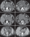
Acute fluid collections in a 52-year-old male patient during the second week of acute necrotizing pancreatitis. A,B,C,D: Contrast-enhanced axial CT images, venous phase showing hypoenhancement of the pancreatic body (arrow on A), compatible with presence of an area of necrosis contiguous with hyperattenuating fluid collection (probable hematic content) in the epiploic retrocavity (arrow on B). Other fluid collections are identified between bowel loops in the peritoneal cavity (long arrows on C, D), in the left anterior pararenal space (short arrow on D), as well as reactive parietal thickening of small loops in the left flank (curved arrow on D) and ascites (black arrow on D).
Figure 5
Figure 5
Disconnected duct syndrome. Acute necrotizing pancreatitis with ductal disconnection in a 61-year-old woman. A,B: Contrast-enhanced axial CT images, parenchymal arterial phase showing area of necrosis in the pancreatic body (long arrow on A) affecting a large portion of the parenchymal thickness, pancreatic tail with preserved appearance (short arrow on A). On B, one identifies the main pancreatic duct discharging into the necrotic area (arrow).
Figure 7
Figure 7
Acute edematous pancreatitis with pseudocysts. A,B: Contrast-enhanced axial CT images, venous phase showing some pseudocysts compressing the pancreatic parenchyma, and others in the epiploic retrocavity (arrows).
Figure 8
Figure 8
Acute edematous pancreatitis with pseudocysts. A,B: Contrast-enhanced axial CT images, venous phase showing preserved enhancement of the pancreatic parenchyma (long arrow on A), pseudocyst posteriorly to the cephalic segment, uncinate process and in the mesenterium (short arrows on A,B).
Figure 9
Figure 9
Pseudocyst in acute pancreatitis. A,B: Contrast-enhanced axial CT image, venous phase showing acute inflammatory changes in the pancreatic tail (long arrows on A) and pseudocyst with spontaneously hyperattenuating hematic content (short arrows on A,B) extending toward the left subphrenic space and partially restrained by the gastric wall (short arrows on A,B).
Figure 10
Figure 10
Post-necrotic pancreatic and peripancreatic changes. A,B: Contrast-enhanced axial CT images, venous phase showing extensive areas of peripancreatic fat necrosis (arrows). C,D: Non-contrast-enhanced CT after eight weeks, such areas become more delimited with a liquefied appearance, characterizing postnecrotic pancreatic and peripancreatic changes (arrows). D,E: A 37-year-old patient with acute necrotizing pancreatitis restricted to peripancreatic tissues. Contrast- enhanced axial CT images, venous phase show preserved pancreatic parenchymal enhancement (long arrows on D,E), with extensive areas of peripancreatic fat necrosis (short arrows on D). The patient presented with a septic condition and was submitted to necrosectomy. Purulent material was identified in those areas.
Figure 11
Figure 11
Walled-off pancreatic necrosis. A,B: Contrast-enhanced axial CT images, venous phase. Development of acute necrotizing pancreatitis in a 45-year-old male patient. A: Extensive necrosis of the pancreatic body and tail with ill-defined limits and solid appearance (arrow). B: After two weeks, the delimitation of the necrotic area with a liquefied appearance can already be observed with necrotic debris inside (arrow). C,D: Contrast-enhanced axial (C) and coronal (D) CT images, venous phase. A 42-year-old male patient with circumscribed parenchymal necrosis replacing the pancreatic body and tail (arrows) after three weeks from the onset of acute necrotizing pancreatitis.
Figure 12
Figure 12
Walled-off pancreatic necrosis. A,B,C: Contrast-enhanced axial CT images, venous phase. Extensive necrosis of the pancreatic body and tail, with peripancreatic inflammatory changes (long arrow on A). Also, a thrombus is identified within the splenic vein (short arrow on A). After approximately one month, an area of walled-off pancreatic necrosis is identified (arrow on B), which should not be confused with pseudocyst. As the same image is evaluated with a narrower window, it is possible to identify the presence of necrotic debris without enhancement within such walled-off pancreatic necrosis (arrows on C). D,E,F: Axial MRI T2-weighted fast spin echo images with (D) and without (E) fat suppression, and non-contrast-enhanced T1-weighted, gradient echo (F) showing necrotic debris of the pancreatic parenchyma deposited in the posterior portion of the collection (arrows on E,F), with signal hyperintensity on T1-weighted sequences, indicating the presence of hemorrhagic component (arrows on E).

References

    1. Vege SS. Clinical manifestations and diagnosis of acute pancreatitis. UpToDate; 2011. [acessado em 20 de setembro de 2011]. Disponível em: https://www.uptodate.com/contents/clinical-manifestations-and-diagnosis-....
    1. Brasil. Ministério da Saúde. Datasus Informações de saúde - 2011. [acessado em 25 de setembro de 2011]. Disponível em: http://tabnet.datasus.gov.br/cgi/tabcgi.exe?sih/cnv/niuf.def.
    1. Bradley EL., 3rd A clinically based classification system for acute pancreatitis. Summary of the International Symposium on Acute Pancreatitis, Atlanta, Ga, September 11 through 13, 1992. Arch Surg. 1993;128:586–590. - PubMed
    1. Bollen TL, van Santvoort HC, Besselink MG, et al. The Atlanta classification of acute pancreatitis revisited. Br J Surg. 2008;95:6–21. - PubMed
    1. Sheu Y, Furlan A, Almusa O, et al. The revised Atlanta classification for acute pancreatitis: a CT imaging guide for radiologists. Emerg Radiol. 2012;19:237–243. - PubMed

LinkOut - more resources