Review
. 2014 Jul-Aug;47(4):228-39.
doi: 10.1590/0100-3984.2013.1762.

Imaging of the adrenal gland lesions

Affiliations
Review

Imaging of the adrenal gland lesions

Keith Herr et al. Radiol Bras. 2014 Jul-Aug.

Abstract

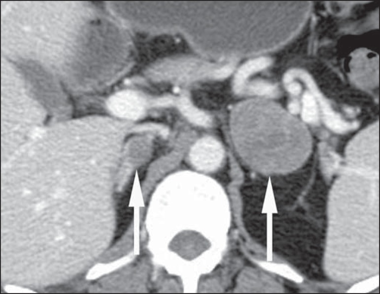
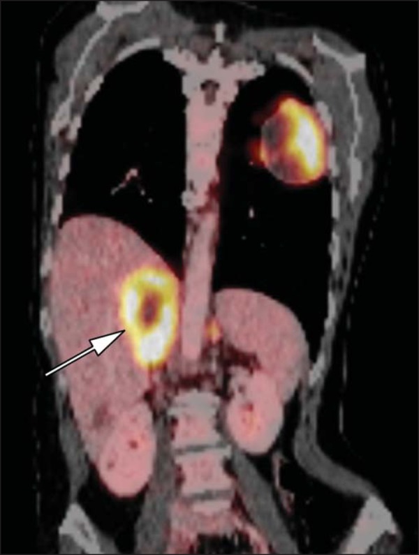
With the steep increase in the use of cross-sectional imaging in recent years, the incidentally detected adrenal lesion, or "incidentaloma", has become an increasingly common diagnostic problem for the radiologist, and a need for an approach to classifying these lesions as benign, malignant or indeterminate with imaging has spurred an explosion of research. While most incidentalomas represent benign disease, typically an adenoma, the possibility of malignant involvement of the adrenal gland necessitates a reliance on imaging to inform management decisions. In this article, we review the literature on adrenal gland imaging, with particular emphasis on computed tomography, magnetic resonance imaging, and photon-emission tomography, and discuss how these findings relate to clinical practice. Emerging technologies, such as contrast-enhanced ultrasonography, dual-energy computed tomography, and magnetic resonance spectroscopic imaging will also be briefly addressed.

O crescente uso da tomografia computadorizada e da ressonância magnética levou a um aumento na identificação de nódulos adrenais incidentais, também chamados de incidentalomas, gerando um impasse diagnóstico para o radiologista, bem como um número significativo de pesquisas a fim de caracterizar essas lesões como benignas ou malignas. Apesar de a maioria dos incidentalomas representar um processo benigno, geralmente um adenoma, a possibilidade de a lesão ser maligna requer suficiente acurácia dos métodos de imagem para que esses possam auxiliar no manejo dos pacientes. Neste artigo nós apresentamos uma revisão da literatura dedicada à investigação radiológica das lesões adrenais, com ênfase na tomografia computadorizada, ressonância magnética e tomografia por emissão de prótons, e discutimos como os achados de imagem relacionam-se com a prática clínica. Tecnologias recentes, como a ultrassonografia com uso de contraste, a tomografia computadorizada com dupla fonte de energia e a espectroscopia de prótons por ressonância magnética são brevemente discutidas.

Keywords: Adenoma; Adrenal gland; Cancer; Diagnosis; Radiology.


Figures

Figure 1
Figure 1
Algorithm for the assessment of an incidental adrenal lesion detected on CT or MRI. (With permission from the American College of Radiology, from Managing incidental findings on abdominal CT: white paper of the ACR incidental findings committee. J Am Coll Radiol. 2010;7:754–73).
Figure 2
Figure 2
Bilateral adrenal adenomas. A region of interest drawn over each adrenal mass measures less than 10 HU, indicative of lipid-rich adenoma.
Figure 3
Figure 3
Incidental adrenal nodule identified on previous CT scan performed for abdominal pain (arrows). A region of interest drawn over an adrenal nodule demonstrated a density of 13 HU on unenhanced CT (A), 80 HU on portal venous phase (B), and 36 HU on the 12-minute delayed phase (C), corresponding to a relative washout of 55% and an absolute washout of 66%. Findings are diagnostic of an adenoma.
Figure 4
Figure 4
Incidental adrenal nodule identified on previous CT scan performed for staging of colon cancer (arrows). The nodule has high signal intensity relative to the spleen on in-phase MR images (A), but this relationship reverses on opposed-phase MR images, as it loses signal from microscopic lipid (B). Findings are diagnostic of a lipid-rich adenoma. S, spleen.
Figure 5
Figure 5
Myelolipoma (arrows). CT demonstrates macroscopic fat-attenuation within the mass (A). On MR imaging, macroscopic fat is characterized by signal loss on fat-suppressed T1-weighted MR images (B, without fat-saturation; C, with fat-saturation, post-contrast).
Figure 6
Figure 6
Adrenal cyst. A small unilocular cyst is identified in the left adrenal gland (arrows). Low signal intensity on T1-weighted images (A), high signal intensity on T2-weighted images (B) similar to the cerebrospinal fluid, and lack of enhancement following the intravenous administration of gadolinium (C) are typical features.
Figure 7
Figure 7
Paracoccidiomycosis. A heterogeneously enhancing adrenal mass is depicted (arrow in A). Paracoccidioides brasiliensis yeast cells in the adrenal gland of a patient with adrenal insufficiency (B) (100× magnification, GMS stain). (With permission from the Instituto de Medicina Tropical de São Paulo, from Identification of Paracoccidioides brasiliensis in adrenal glands biopsies of two patients with paracoccidiomycosis and adrenal insufficiency. Rev Inst Med Trop S Paulo. 2009;51:45–8).
Figure 8
Figure 8
Hematoma. A: A focal, non-enhancing, homogenous right adrenal mass measuring greater than water attenuation (30 HU) on contrast-enhanced CT in a patient with Merkel cell carcinoma as part of PET-CT examination for surveillance (arrow). PET portion demonstrated a corresponding area of photopenia (not shown). B: An unenhanced CT scan performed three months earlier demonstrates a normal right adrenal gland (arrow).
Figure 9
Figure 9
Pheochromocytoma (arrows). A large, heterogeneous adrenal mass on a T2-weighted MR image (A). Cystic and solid enhancing components are depicted in a post-contrast MR image (B). Elevated serum catecholamines were detected in this patient with pheochromocytoma.
Figure 10
Figure 10
Pheochromocytoma (arrows). A contrast-enhanced CT image demonstrates a somewhat heterogeneously enhancing adrenal mass (A). Focal increased radiotracer uptake (B) corresponding to the adrenal mass in (A) on coronal 123-I MIBG scintigraphy. Physiologic radiotracer activity in the liver and excretion in the urinary bladder are noted.
Figure 11
Figure 11
Adrenocortical carcinoma. A heterogeneously enhancing adrenal mass (asterisk) is demonstrated, which displaces the ipsilateral kidney laterally. This mass was diagnosed as adrenocortical carcinoma at biopsy. Note invasion of the left renal vein (arrowhead). IVC, inferior vena cava.
Figure 12
Figure 12
Neuroblastoma. A large, heterogeneously enhancing adrenal mass (asterisk) with encasement of the celiac axis (arrowhead) and other abdominal vessels is shown. Calcifications are common, but not present in this case. S, spleen.
Figure 13
Figure 13
Melanoma metastases. Post-contrast image demonstrates bilateral adrenal masses (arrows). While the imaging features of these lesions are not specific, the bilateral distribution, size of the left-sided mass, and history of melanoma suggest the diagnosis.
Figure 14
Figure 14
Lung cancer metastasis. Coronal fused FDG-PET/CT with a large, hypermetabolic right adrenal mass with central necrosis, indicative of metastatic disease (arrow) in this patient.

Similar articles

Cited by

References

    1. Zeiger MA, Siegelman SS, Hamrahian AH. Medical and surgical evaluation and treatment of adrenal incidentalomas. J Clin Endocrinol Metab. 2011;96:2004–2015. - PubMed
    1. Young WF., Jr Clinical practice. The incidentally discovered adrenal mass. N Engl J Med. 2007;356:601–610. - PubMed
    1. Song JH, Chaudhry FS, Mayo-Smith WW. The incidental adrenal mass on CT: prevalence of adrenal disease in 1,049 consecutive adrenal masses in patients with no known malignancy. AJR Am J Roentgenol. 2008;190:1163–1168. - PubMed
    1. Lenert JT, Barnett CC, Jr, Kudelka AP, et al. Evaluation and surgical resection of adrenal masses in patients with a history of extraadrenal malignancy. Surgery. 2001;130:1060–1067. - PubMed
    1. Lam KY, Lo CY. Metastatic tumours of the adrenal glands: a 30-year experience in a teaching hospital. Clin Endocrinol. 2002;56:95–101. - PubMed

LinkOut - more resources