

Urachal neoplasia: a case report
- PMID: 25741124
- PMCID: PMC4341378
- DOI: 10.1590/0100-3984.2013.1879
Urachal neoplasia: a case report
Abstract
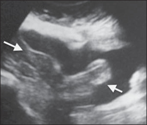
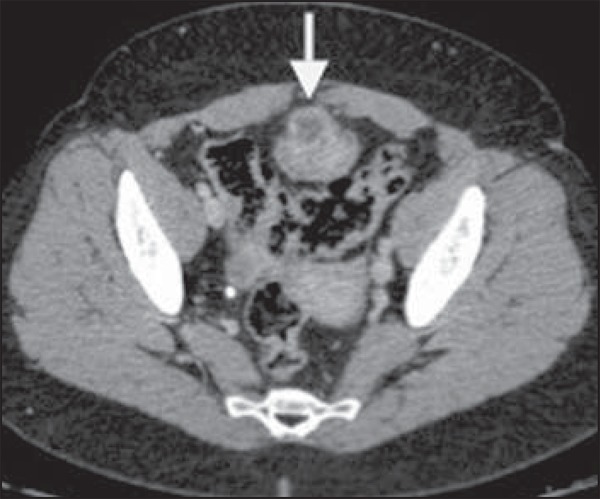
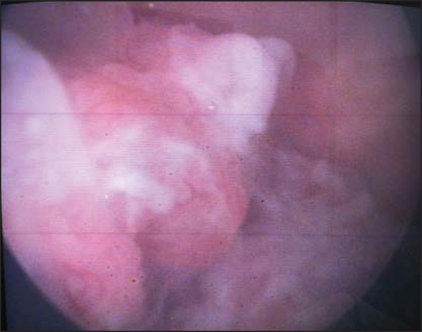
Diseases of urachal remnants are uncommon and generally located on the junction of the urachal remnant with the bladder dome. In most cases such diseases correspond to mucinous adenocarcinomas and present hematuria as their most common clinical finding. The authors report the case of a 62-year-old female patient undergoing follow-up due to macroscopic hematuria for three years. Ultrasonography and computed tomography findings are described.
As doenças dos remanescentes do úraco são incomuns. Localizam-se geralmente na junção remanescente do úraco com a cúpula vesical. Adenocarcinomas mucinosos são a maioria. A característica clínica mais comum é a hematúria. Neste artigo relatamos o caso de uma paciente de 62 anos com quadro de hematúria macroscópica com evolução de três anos. São descritos achados na ultrassonografia e tomografia computadorizada.
Keywords: Carcinoma; Hematuria; Neoplasms of the bladder; Urachus.
Figures

References
-
- Yu JS, Kim KW, Lee HJ, et al. Urachal remnant diseases: spectrum of CT and US findings. Radiographics. 2001;21:451–461. - PubMed
-
- Koster IM, Cleyndert P, Giard RW. Best cases from the AFIP: urachal carcinoma. Radiographics. 2009;29:939–942. - PubMed
-
- Ashley RA, Inman BA, Sebo TJ, et al. Urachal carcinoma: clinicopathologic features and long-term outcomes of an aggressive malignancy. Cancer. 2006;107:712–720. - PubMed
-
- Donate Moreno MJ, Giménez Bachs JM, Salinas Sánchez AS, et al. Patología del uraco: revisión de conjunto y presentación de tres casos. Actas Urol Esp. 2005;29:332–336. - PubMed
-
- Thali-Schwab CM, Woodward PJ, Wagner BJ. Computed tomographic appearance of urachal adenocarcinomas: review of 25 cases. Eur Radiol. 2005;15:79–84. - PubMed
Publication types
LinkOut - more resources
Full Text Sources
Other Literature Sources