

Diagnosis of ectopic pancreas by endoscopic ultrasound with fine-needle aspiration
- PMID: 25741143
- PMCID: PMC4342912
- DOI: 10.3748/wjg.v21.i8.2367
Diagnosis of ectopic pancreas by endoscopic ultrasound with fine-needle aspiration
Abstract
Aim: To study the clinical, endoscopic, sonographic, and cytologic features of ectopic pancreas (EP).
Methods: This was a retrospective study performed at an academic referral center including two hospitals. Institutional review board approval was obtained. Patients referred to the University Hospital or Denver Health Medical Center Gastrointestinal Endoscopy Lab for gastroduodenal subepithelial lesions (SEL) with a final diagnosis of EP between January 2009 and December 2013 were identified. Patients in this group were selected for the study if they underwent endoscopic ultrasound (EUS) with fine-needle aspiration (FNA) or deep biopsy. A review of the medical record was performed specifically to review the following information: presenting symptoms, endoscopic and EUS findings, computed tomography or magnetic resonance imaging findings, pathology results, procedure-related adverse events, and subsequent treatments after EUS-FNA. EUS with FNA or deep submucosal biopsy was performed in all patients on an outpatient basais by one of two physicians (Attwell A, Fukami N). Review of all subsequent clinic notes and operative reports was performed in order to determine follow-up and final diagnoses.
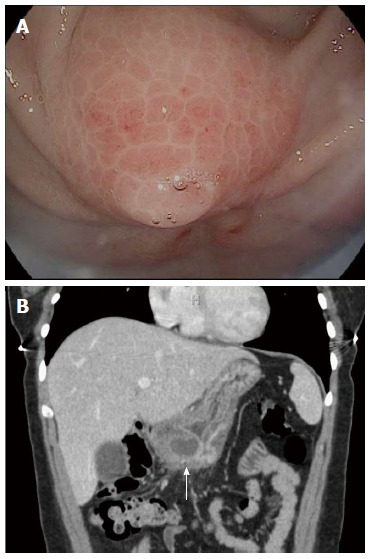
Results: Between July 2009 and December 2013, 10 patients [3 males, 7 females, median age 52 (26-64) years] underwent EUS for a gastroduodenal SEL and were diagnosed with EP. One patient was symptomatic. Six (60%) lesions were in the antrum, 3 (30%) in the body, and 1 (10%) in the duodenum. A mucosal dimple was noted in 6 (60%). Mean lesion size was 17 (8-25) mm. Gastrointestinal wall involvement: muscularis mucosae, 10%; submucosa, 70%; muscularis propria, 60%; and serosa, 10%. Nine (90%) lesions were hypoechoic and 5 (50%) were homogenous. A duct was seen in 5 (50%). FNA was attempted in 9 (90%) and successful in 8 (80%) patients after 4 (2-6) passes. Cytology showed acini or ducts in 7 of 8 (88%). Superficial biopsies in 7 patients (70%) showed normal gastric mucosa. Deep endoscopic biopsies were taken in 2 patients and diagnostic in one. One patient (10%) developed pancreatitis after EUS-FNA. Two patients (20%) underwent surgery to relieve symptoms or confirm the diagnosis. The main limitation of the study was the fact that it was retrospective and performed at a single medical center.
Conclusion: EUS features of EP include antral location, mucosal dimple, location in layers 3-4, and lesional duct, and FNA or biopsy is accurate and effective.
Keywords: Ectopic pancreas; Endoscopic ultrasound; Endoscopy; Pancreatitis; Subepithelial mass.
Figures


Similar articles
-
Lesion size determines diagnostic yield of EUS-FNA with onsite cytopathologic evaluation for upper gastrointestinal subepithelial lesions.Turk J Gastroenterol. 2018 Jul;29(4):436-441. doi: 10.5152/tjg.2018.17876. Turk J Gastroenterol. 2018. PMID: 30249558 Free PMC article.
-
Clinical usefulness of endoscopic ultrasound-guided fine needle aspiration for gastric subepithelial lesions smaller than 2 cm.J Gastrointestin Liver Dis. 2014 Dec;23(4):405-12. doi: 10.15403/jgld.2014.1121.234.eug. J Gastrointestin Liver Dis. 2014. PMID: 25531999
-
Factors affecting the diagnostic accuracy of endoscopic ultrasonography-guided fine-needle aspiration (EUS-FNA) for upper gastrointestinal submucosal or extraluminal solid mass lesions.Dig Endosc. 2012 Sep;24(5):358-63. doi: 10.1111/j.1443-1661.2012.01243.x. Epub 2012 Mar 13. Dig Endosc. 2012. PMID: 22925290
-
Pretherapeutic evaluation of patients with upper gastrointestinal tract cancer using endoscopic and laparoscopic ultrasonography.Dan Med J. 2012 Dec;59(12):B4568. Dan Med J. 2012. PMID: 23290296 Review.
-
Endoscopic submucosal dissection combined surgery for the treatment of ectopic gastric mucosa and ectopic pancreas in muscularis propria and serosal layer of the stomach: A rare case report and review of the literature.Medicine (Baltimore). 2025 Feb 28;104(9):e41297. doi: 10.1097/MD.0000000000041297. Medicine (Baltimore). 2025. PMID: 40020126 Free PMC article. Review.
Cited by
-
Ectopic pancreas in the upper gastrointestinal tract: Is endosonographic diagnosis reliable? Data from the German Endoscopic Ultrasound Registry and review of the literature.Endosc Ultrasound. 2018 Jul-Aug;7(4):270-278. doi: 10.4103/eus.eus_18_17. Endosc Ultrasound. 2018. PMID: 28836514 Free PMC article.
-
Evaluation and management of a pancreatic rest noted during pre-bariatric surgery screening endoscopy.Surg Endosc. 2021 Feb;35(2):536-561. doi: 10.1007/s00464-020-08040-2. Epub 2020 Oct 1. Surg Endosc. 2021. PMID: 33006030 Review.
-
The impact of age and sex on the occurrence of pathology in the wall of the upper gastrointestinal tract.Prz Gastroenterol. 2017;12(3):192-198. doi: 10.5114/pg.2017.70472. Epub 2017 Sep 30. Prz Gastroenterol. 2017. PMID: 29123580 Free PMC article.
-
Various Phenotypes of Ectopic Pancreatic Tissue in Children: Case Series and Literature Review.Diagnostics (Basel). 2025 May 8;15(10):1193. doi: 10.3390/diagnostics15101193. Diagnostics (Basel). 2025. PMID: 40428186 Free PMC article.
-
Interventional endoscopic ultrasound for a symptomatic pseudocyst secondary to gastric heterotopic pancreas.World J Gastroenterol. 2017 Sep 14;23(34):6365-6370. doi: 10.3748/wjg.v23.i34.6365. World J Gastroenterol. 2017. PMID: 28974904 Free PMC article.
References
-
- Trifan A, Târcoveanu E, Danciu M, Huţanaşu C, Cojocariu C, Stanciu C. Gastric heterotopic pancreas: an unusual case and review of the literature. J Gastrointestin Liver Dis. 2012;21:209–212. - PubMed
-
- Rösch T, Kapfer B, Will U, Baronius W, Strobel M, Lorenz R, Ulm K. Accuracy of endoscopic ultrasonography in upper gastrointestinal submucosal lesions: a prospective multicenter study. Scand J Gastroenterol. 2002;37:856–862. - PubMed
-
- Karaca C, Turner BG, Cizginer S, Forcione D, Brugge W. Accuracy of EUS in the evaluation of small gastric subepithelial lesions. Gastrointest Endosc. 2010;71:722–727. - PubMed
-
- Matsushita M, Hajiro K, Okazaki K, Takakuwa H. Gastric aberrant pancreas: EUS analysis in comparison with the histology. Gastrointest Endosc. 1999;49:493–497. - PubMed
-
- Changchien CS, Hsiaw CM, Hu TH. Endoscopic ultrasonographic classification of gastric aberrant pancreas. Chang Gung Med J. 2000;23:600–607. - PubMed
MeSH terms
LinkOut - more resources
Full Text Sources
Other Literature Sources
Medical
Miscellaneous
