

Characterization of breast lesions: comparison of digital breast tomosynthesis and ultrasonography
- PMID: 25741187
- PMCID: PMC4347261
- DOI: 10.3348/kjr.2015.16.2.229
Characterization of breast lesions: comparison of digital breast tomosynthesis and ultrasonography
Abstract
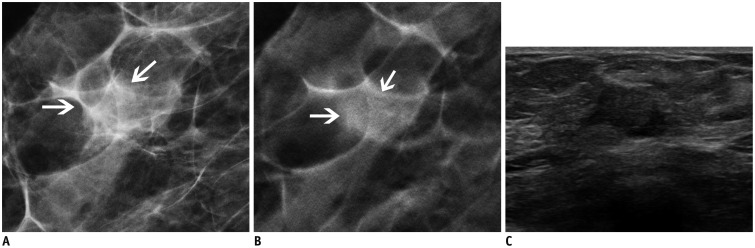
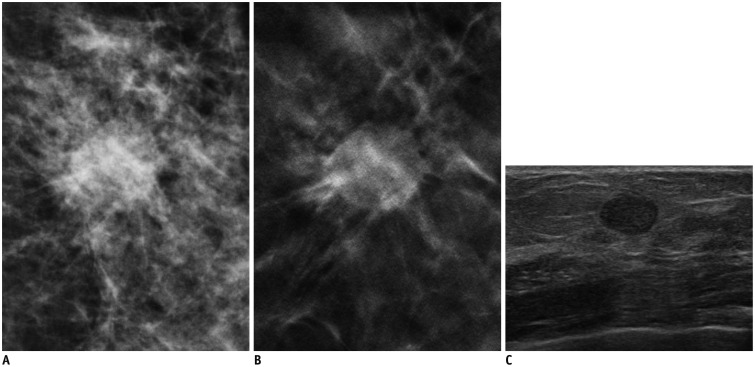
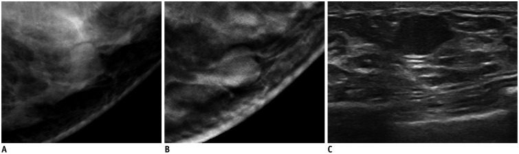
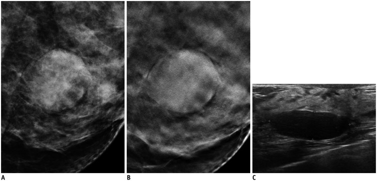
Objective: To compare the diagnostic performance of digital breast tomosynthesis (DBT) and conventional breast ultrasound (US) to characterize breast lesions as benign or malignant.
Materials and methods: A total of 332 women, presenting for screening examinations or for breast biopsy between March and June 2012 were recruited to undergo digital mammography (DM), DBT, and breast US examination. Among them, 113 patients with 119 breast lesions depicted on DM were finally included. Three blinded radiologists performed an enriched reader study and reviewed the DBT and US images. Each reader analyzed the lesions in random order, assigned Breast Imaging Reporting and Data System (BI-RADS) descriptors, rated the images for the likelihood of malignancy (%) and made a BI-RADS final assessment. Diagnostic accuracy, as assessed by the area under the receiver operating characteristic curve, sensitivity, and specificity of DBT and US were compared.
Results: Among the 119 breast lesions depicted on DM, 75 were malignant and the remaining 44 were benign. The average diagnostic performance for characterizing breast lesions as benign or malignant in terms of area under the curve was 0.899 for DBT and 0.914 for US (p = 0.394). Mean sensitivity (97.3% vs. 98.7%, p = 0.508) and specificity (44.7% vs. 39.4%, p = 0.360) were also not significantly different.
Conclusion: Digital breast tomosynthesis may provide similar reader lesion characterization performance to that of US for breast lesions depicted on DM.
Keywords: Breast US; Diagnostic performance; Digital breast tomosynthesis.
Figures
Similar articles
-
Characterization of Breast Masses in Digital Breast Tomosynthesis and Digital Mammograms: An Observer Performance Study.Acad Radiol. 2017 Nov;24(11):1372-1379. doi: 10.1016/j.acra.2017.04.016. Epub 2017 Jun 21. Acad Radiol. 2017. PMID: 28647388 Free PMC article.
-
Additional US or DBT after digital mammography: which one is the best combination?Acta Radiol. 2016 Jan;57(1):13-8. doi: 10.1177/0284185114563641. Epub 2014 Dec 18. Acta Radiol. 2016. PMID: 25523063
-
Digital breast tomosynthesis and breast ultrasound: Additional roles in dense breasts with category 0 at conventional digital mammography.Eur J Radiol. 2016 Jan;85(1):291-296. doi: 10.1016/j.ejrad.2015.09.026. Epub 2015 Sep 30. Eur J Radiol. 2016. PMID: 26499000
-
Digital breast tomosynthesis in the diagnostic environment: A subjective side-by-side review.AJR Am J Roentgenol. 2010 Aug;195(2):W172-6. doi: 10.2214/AJR.09.3244. AJR Am J Roentgenol. 2010. PMID: 20651178 Review.
-
Accuracy of Digital Breast Tomosynthesis for Detecting Breast Cancer in the Diagnostic Setting: A Systematic Review and Meta-Analysis.Korean J Radiol. 2021 Aug;22(8):1240-1252. doi: 10.3348/kjr.2020.1227. Epub 2021 May 20. Korean J Radiol. 2021. PMID: 34047504 Free PMC article.
Cited by
-
Associating Automated Breast Ultrasound (ABUS) and Digital Breast Tomosynthesis (DBT) with Full-Field Digital Mammography (FFDM) in Clinical Practice in Cases of Women with Dense Breast Tissue.Diagnostics (Basel). 2022 Feb 11;12(2):459. doi: 10.3390/diagnostics12020459. Diagnostics (Basel). 2022. PMID: 35204550 Free PMC article.
-
Selection and Reporting of Statistical Methods to Assess Reliability of a Diagnostic Test: Conformity to Recommended Methods in a Peer-Reviewed Journal.Korean J Radiol. 2017 Nov-Dec;18(6):888-897. doi: 10.3348/kjr.2017.18.6.888. Epub 2017 Sep 21. Korean J Radiol. 2017. PMID: 29089821 Free PMC article.
-
Performance of Screening Mammography: A Report of the Alliance for Breast Cancer Screening in Korea.Korean J Radiol. 2016 Jul-Aug;17(4):489-96. doi: 10.3348/kjr.2016.17.4.489. Epub 2016 Jun 27. Korean J Radiol. 2016. PMID: 27390540 Free PMC article.
-
Analysis of Participant Factors That Affect the Diagnostic Performance of Screening Mammography: A Report of the Alliance for Breast Cancer Screening in Korea.Korean J Radiol. 2017 Jul-Aug;18(4):624-631. doi: 10.3348/kjr.2017.18.4.624. Epub 2017 May 19. Korean J Radiol. 2017. PMID: 28670157 Free PMC article.
-
Addition of Digital Breast Tomosynthesis to Full-Field Digital Mammography in the Diagnostic Setting: Additional Value and Cancer Detectability.J Breast Cancer. 2016 Dec;19(4):438-446. doi: 10.4048/jbc.2016.19.4.438. Epub 2016 Dec 23. J Breast Cancer. 2016. PMID: 28053633 Free PMC article.
References
-
- Berry DA, Cronin KA, Plevritis SK, Fryback DG, Clarke L, Zelen M, et al. Effect of screening and adjuvant therapy on mortality from breast cancer. N Engl J Med. 2005;353:1784–1792. - PubMed
-
- Hellquist BN, Duffy SW, Abdsaleh S, Björneld L, Bordás P, Tabár L, et al. Effectiveness of population-based service screening with mammography for women ages 40 to 49 years: evaluation of the Swedish Mammography Screening in Young Women (SCRY) cohort. Cancer. 2011;117:714–722. - PubMed
-
- Tabár L, Fagerberg CJ, Gad A, Baldetorp L, Holmberg LH, Gröntoft O, et al. Reduction in mortality from breast cancer after mass screening with mammography. Randomised trial from the Breast Cancer Screening Working Group of the Swedish National Board of Health and Welfare. Lancet. 1985;1:829–883. - PubMed
-
- Tabar L, Yen MF, Vitak B, Chen HH, Smith RA, Duffy SW. Mammography service screening and mortality in breast cancer patients: 20-year follow-up before and after introduction of screening. Lancet. 2003;361:1405–1410. - PubMed
-
- Mandelson MT, Oestreicher N, Porter PL, White D, Finder CA, Taplin SH, et al. Breast density as a predictor of mammographic detection: comparison of interval- and screen-detected cancers. J Natl Cancer Inst. 2000;92:1081–1087. - PubMed
Publication types
MeSH terms
LinkOut - more resources
Full Text Sources
Other Literature Sources
Medical
