Review
. 2015 Mar-Apr;16(2):239-50.
doi: 10.3348/kjr.2015.16.2.239. Epub 2015 Feb 27.

Coronary artery imaging in children

Affiliations
Review

Coronary artery imaging in children

Hyun Woo Goo. Korean J Radiol. 2015 Mar-Apr.

Abstract

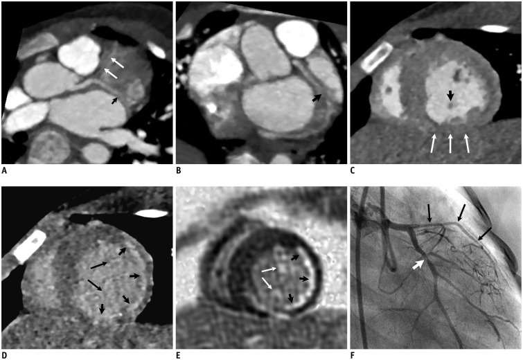
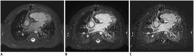
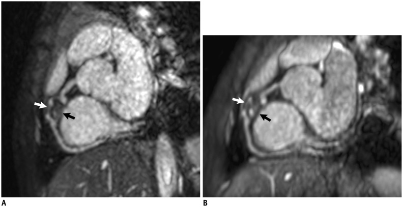
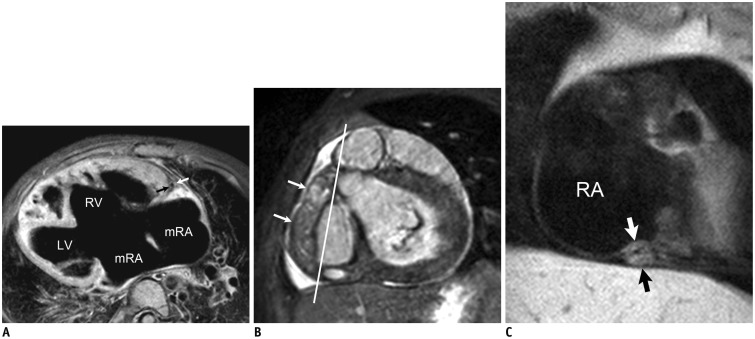
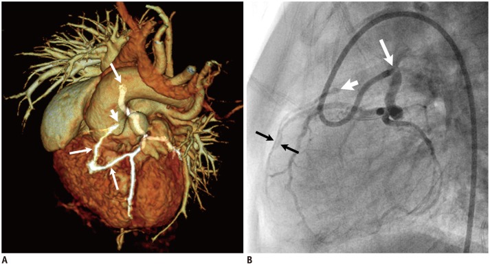
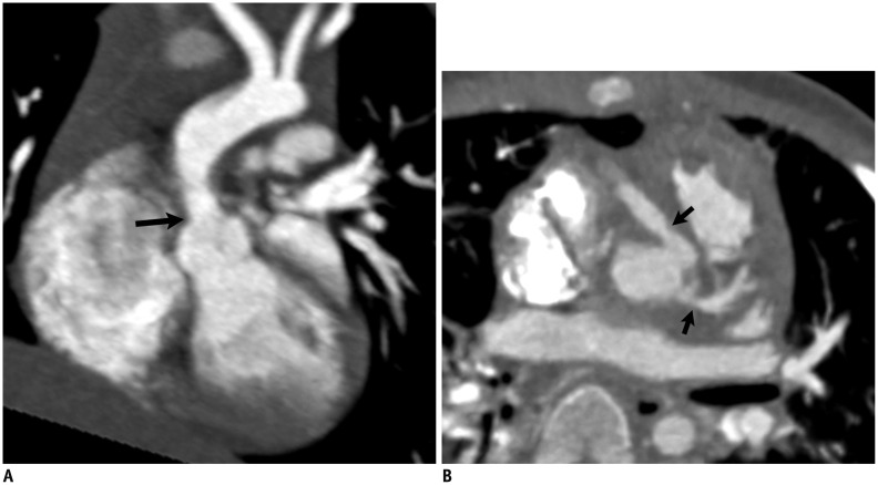
Coronary artery problems in children usually have a significant impact on both short-term and long-term outcomes. Early and accurate diagnosis, therefore, is crucial but technically challenging due to the small size of the coronary artery, high heart rates, and limited cooperation of children. Coronary artery visibility on CT and MRI in children is considerably improved with recent technical advancements. Consequently, CT and MRI are increasingly used for evaluating various congenital and acquired coronary artery abnormalities in children, such as coronary artery anomalies, aberrant coronary artery anatomy specific to congenital heart disease, Kawasaki disease, Williams syndrome, and cardiac allograft vasculopathy.

Keywords: CT; Catheter angiography; Coronary artery; Echocardiography; Infants and children; MRI.


Figures

Fig. 1
Fig. 1. 3-month-old boy with anomalous origin of left coronary artery (LCA) from pulmonary artery.
Transthoracic echocardiographic images (A, B) show normal origins of right coronary artery (RCA, arrow in A) and LCA (arrow in B). C. Oblique axial cardiac CT image demonstrates normal origin of RCA (arrowhead) but no connection (arrow) of LCA to left aortic sinus. D. Cropped oblique coronal volume-rendered CT image clearly reveals anomalous origin (black arrow) of LCA from pulmonary trunk (PA) (white arrow). Dilated left ventricle (LV) is noted. AO = ascending aorta, RVOT = right ventricular outflow tract
Fig. 2
Fig. 2. 11-year-old boy who underwent double switch operation for congenitally-corrected TGA.
A. 4-chamber cardiac CT image shows morphologic left ventricle (LV), morphologic right ventricle (RV), morphologic right atrium (RA), morphologic left atrium (LA), and patent systemic venous baffle (asterisks). Oblique axial (B-D) and coronal (E) cardiac CT images demonstrate patent transferred coronary arteries, dual RCA (RCA1, RCA2), and patent systemic venous baffle (asterisks). IVC = inferior vena cava, RCA = right coronary artery, SVC = superior vena cava, TGA = transposition of great arteries
Fig. 3
Fig. 3. 3-year-old boy with cardiac involvement of chronic graft-versus-host disease after hematopoietic stem cell transplantation for chronic granulomatous disease.
Cardiac CT images (A, B) show long-segment severe narrowing (long arrows) of left anterior descending artery and focal narrowing (short arrow) of proximal left circumflex artery. C. Early short-axis cardiac CT image reveals thinning of inferior wall (long arrows) and posteromedial papillary muscle (short arrow) of left ventricle. D. 6-min-delayed short-axis cardiac CT image demonstrates subendocardial myocardial delayed enhancement (short arrows) in left coronary artery territory and left ventricular papillary muscles (long arrows) indicating myocardial infarction, which is in accordance with short-axis late gadolinium enhancement MR image (E). F. Catheter left coronary angiography confirms long-segment severe narrowing (long arrows) of left anterior descending artery and focal narrowing (short arrow) of proximal left circumflex artery as seen on cardiac CT (A, B).
Fig. 4
Fig. 4. 3-year-old boy with functional single ventricle who underwent extracardiac conduit Fontan operation.
Pre- (A) and post-contrast (B) navigator-gated coronary MR angiographic images obtained at 1.5-T MR scanner demonstrate that cardiovascular structures are substantially enhanced after intravenous administration of extracellular gadolinium-based contrast agent. C. Subtraction image clearly show effect of contrast-enhancement in coronary MR angiography. Normal enhancement of dependent portions of both lungs is also noted (B, C).
Fig. 5
Fig. 5. 6-year-old boy with Kawasaki disease.
Target-volume (A) and whole-heart (B) navigator-gated coronary MR angiographic images show large proximal right coronary artery aneurysm (arrows) with thrombus with comparable image quality.
Fig. 6
Fig. 6. Coronary vessel wall MRI.
A. Axial electrocardiography (ECG)-triggered, navigator-gated, double inversion recovery, fat-saturated black-blood segmented turbo spin-echo MRI in 3-year-old girl with functional single ventricle and right isomerism who underwent bidirectional cavopulmonary shunt shows wall (arrows) of normal right coronary artery. In addition, dextrocardia, large ventricular septal defect, and remnant of interatrial septum are noted. C. Oblique coronal ECG-triggered, navigator-gated, double inversion recovery, fat-saturated black-blood segmented turbo spin-echo MRI obtained along line in B in 3-year-old boy with Kawasaki disease and thrombosed fusiform aneurysm (long arrows in B) of right coronary artery reveals severe concentric wall thickening (short arrows in C) of distal right coronary artery. LV = left ventricle, mRA = morphologic right atrium, RA = right atrium, RV = right ventricle
Fig. 7
Fig. 7. 9-month-old girl with tetralogy of Fallot who underwent left Blalock-Taussig shunt.
Volume-rendered cardiac CT image (A) and lateral view of catheter left coronary angiography (B) demonstrate high take-off (long arrow) left coronary artery, atresia (short arrow) of right coronary artery, and collateral artery between left and right coronary arteries (long arrows).
Fig. 8
Fig. 8. 5-month-old girl who underwent reimplantation of anomalous left coronary artery from pulmonary artery.
A. Cropped coronal volume-rendered cardiac CT image shows extrinsic compression (arrow) of reimplanted left main coronary artery for pulmonary trunk (PT) that is better depicted on segmented volume-rendered image of coronary artery and pulmonary artery (B).
Fig. 9
Fig. 9. 10-month-old boy with Williams syndrome.
A. Oblique coronal cardiac CT image shows supravavular aortic stenosis (arrow). B. Oblique axial cardiac CT image reveals ostial stenoses (arrows) of left and right coronary arteries.

Similar articles

Cited by

References

    1. Lederlin M, Thambo JB, Latrabe V, Corneloup O, Cochet H, Montaudon M, et al. Coronary imaging techniques with emphasis on CT and MRI. Pediatr Radiol. 2011;41:1516–1525. - PubMed
    1. Goo HW. State-of-the-art CT imaging techniques for congenital heart disease. Korean J Radiol. 2010;11:4–18. - PMC - PubMed
    1. Tang L, Merkle N, Schär M, Korosoglou G, Solaiyappan M, Hombach V, et al. Volume-targeted and whole-heart coronary magnetic resonance angiography using an intravascular contrast agent. J Magn Reson Imaging. 2009;30:1191–1196. - PubMed
    1. Vranicar M, Hirsch R, Canter CE, Balzer DT. Selective coronary angiography in pediatric patients. Pediatr Cardiol. 2000;21:285–288. - PubMed
    1. Attili A, Hensley AK, Jones FD, Grabham J, DiSessa TG. Echocardiography and coronary CT angiography imaging of variations in coronary anatomy and coronary abnormalities in athletic children: detection of coronary abnormalities that create a risk for sudden death. Echocardiography. 2013;30:225–233. - PubMed

MeSH terms