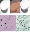
Single nodular opacity of granulomatous pneumocystis jirovecii pneumonia in an asymptomatic lymphoma patient
- PMID: 25741206
- PMCID: PMC4347280
- DOI: 10.3348/kjr.2015.16.2.440
Single nodular opacity of granulomatous pneumocystis jirovecii pneumonia in an asymptomatic lymphoma patient
Abstract
The radiologic findings of a single nodule from Pneumocystis jirovecii pneumonia (PJP) have been rarely reported. We described a case of granulomatous PJP manifesting as a solitary pulmonary nodule with a halo sign in a 69-year-old woman with diffuse large B cell lymphoma during chemotherapy. The radiologic appearance of the patient suggested an infectious lesion such as angioinvasive pulmonary aspergillosis or lymphoma involvement of the lung; however, clinical manifestations were not compatible with the diseases. The nodule was confirmed as granulomatous PJP by video-assisted thoracoscopic surgery biopsy.
Keywords: Granulomatous Pneumocystis jirovecii pneumonia; Lymphoma; Nodular opacity.
Figures

References
-
- Boiselle PM, Crans CA, Jr, Kaplan MA. The changing face of Pneumocystis carinii pneumonia in AIDS patients. AJR Am J Roentgenol. 1999;172:1301–1309. - PubMed
-
- Marchiori E, Müller NL, Soares Souza A, Jr, Escuissato DL, Gasparetto EL, Franquet T. Pulmonary disease in patients with AIDS: high-resolution CT and pathologic findings. AJR Am J Roentgenol. 2005;184:757–764. - PubMed
-
- Pastores SM, Garay SM, Naidich DP, Rom WN. Review: pneumothorax in patients with AIDS-related Pneumocystis carinii pneumonia. Am J Med Sci. 1996;312:229–234. - PubMed
-
- Hartz JW, Geisinger KR, Scharyj M, Muss HB. Granulomatous pneumocystosis presenting as a solitary pulmonary nodule. Arch Pathol Lab Med. 1985;109:466–469. - PubMed
-
- Chang H, Shih LY, Wang CW, Chuang WY, Chen CC. Granulomatous Pneumocystis jiroveci pneumonia in a patient with diffuse large B-cell lymphoma: case report and review of the literature. Acta Haematol. 2010;123:30–33. - PubMed
Publication types
MeSH terms
Substances
LinkOut - more resources
Full Text Sources
Other Literature Sources

