Review
. 2015;3(1):39-55.
doi: 10.1007/s40336-014-0098-3. Epub 2015 Jan 21.

Amyloid PET imaging: applications beyond Alzheimer's disease

Affiliations
Review

Amyloid PET imaging: applications beyond Alzheimer's disease

Ana M Catafau et al. Clin Transl Imaging. 2015.

Abstract

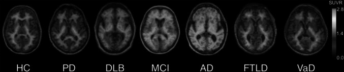
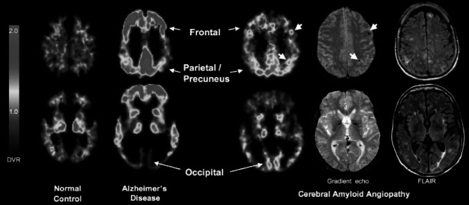
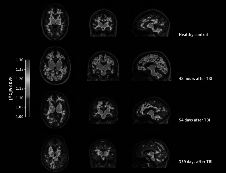
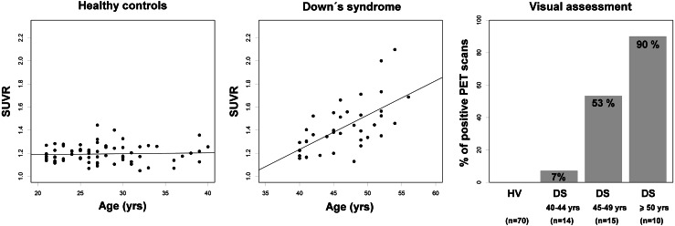
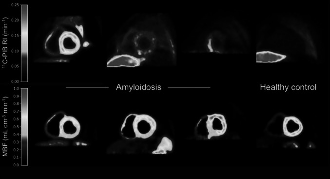
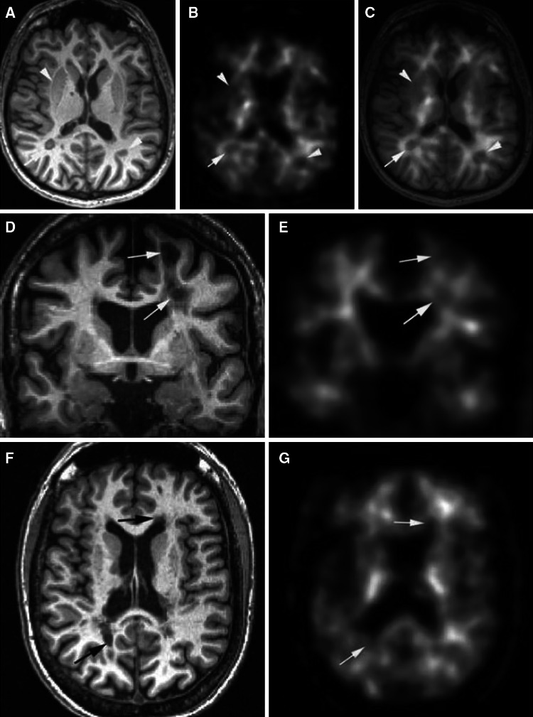
As a biomarker of beta-amyloid, positron emission tomography (PET) amyloid imaging offers a unique opportunity to detect the presence of this protein in the human body during life. Besides Alzheimer's disease (AD), deposits of beta-amyloid in the brain are also present in other neurodegenerative diseases associated to dementia, such as Parkinson's disease and dementia with Lewy bodies, as well as in other processes affecting brain function, such as cerebral amyloid angiopathy, brain trauma, Down's syndrome and meningiomas, as shown by post-mortem pathology studies. Furthermore, in systemic amyloidosis other organs besides the brain are affected, and amyloid PET imaging may be suitable for the identification of these extra-cerebral amyloid depositions. Finally, the potential use of amyloid PET tracer accumulation in cerebral white matter (WM) as a marker of myelin is being investigated, leading to some promising results in patients with WM lesions and multiple sclerosis. In this article, a review of the ongoing research pointing to a broader application of amyloid PET imaging in clinical practice beyond AD is provided.

Keywords: Amyloid PET; Brain trauma; Cardiac amyloidosis; Cerebral amyloid angiopathy; Down’s syndrome; Multiple sclerosis.


Figures

Fig. 1
Fig. 1
Representative 18F-florbetaben PET transaxial images overlaid on individual coregistered MR images of a healthy control (HC) and patients with Parkinson’s disease (PD), dementia with Lewy bodies (DLB), mild cognitive impairment (MCI), Alzheimer’s disease (AD), frontotemporal lobar degeneration (FTLD), and vascular dementia (VaD). Only the DLB, MCI and AD patients show cortical 18F-florbetaben uptake in addition to the non-specific white matter uptake visible in negative scans of HC, PD, FTLD, and VaD. The cortical tracer uptake in DLB is lower than in AD. All images are scaled to same SUVR maximum. This research was originally published in JNM. Villemagne et al. [11] © by the Society of Nuclear Medicine and Molecular Imaging, Inc
Fig. 2
Fig. 2
Representative 11C-PIB PET images at two transaxial levels from normal control (NC) (11C-PIB-negative), Alzheimer’s disease (AD), and cerebral amyloid angiopathy (CAA) subjects. Compared with AD and NC, CAA subjects had an intermediate level of global 11C-PIB retention, but compared with AD, had relatively increased occipital retention. Microbleeds seen in this patient, shown in coregistered gradient echo magnetic resonance images, at times appear proximal to foci of amyloid deposition (small arrows). Reproduced from Ann Neurol, Johnson et al. [34] ©2007 with permission from the American Neurological Association
Fig. 3
Fig. 3
11C-PIB distribution volume ratio (DVR) maps from a control and three patients scanned at different times after TBI. Note the progressive decrease in 11C-PIB uptake as time after TBI increases. Reproduced from JAMA Neurol, Hong YT et al. (doi:10.1001/jamaneurol.2013.4847) with permission from American Medical Association
Fig. 4
Fig. 4
18F-florbetaben SUVR versus age in young cognitively normal healthy controls (n = 70; SUVR = 0.001 × age + 1.173, R 2 = −0.01) (left graph) (Piramal Imaging, unpublished data) and subjects with Down’s syndrome (n = 39; SUVR = 0.030 × age + 0.045, R 2 = 0.39) (middle graph). A graph showing the percentage of positive 18F-florbetaben scans grouped by age clusters is shown in the right hand side
Fig. 5
Fig. 5
Short-axis images of 11C-PIB retention index (RI) and myocardial blood flow (MBF) in (left to right) patients with high, intermediate, and partially increased 11C-PIB retention and a healthy control. Liver is clearly visible in 11C-PIB images of one patient (second column images) patient and healthy control, and is just outside PET field of view for other two patients. Liver uptake is due to biliary excretion of 11C-PIB and is likely not related to amyloid binding. This research was originally published in JNM. Antoni et al. [78] © by the Society of Nuclear Medicine and Molecular Imaging, Inc
Fig. 6
Fig. 6
MP-RAGE 3T MRI (a, d, f) and corresponding co-registered 11C-PIB slices (b, c, e, g) from two representative patients from Stankoff et al. [83] (dg correspond to the same patient). c Shows superposition of MRI and PET images. Arrows point to MA plaques, and arrowheads point to gray matter structures, both appearing as a loss of uptake on PET images. Reproduced from Ann Neurol, Stankoff et al. [83] with permission from the American Neurological Association

Similar articles

Cited by

References

    1. Villemagne VL, Mulligan RS, Pejoska S, Ong K, Jones G, O’Keefe G, Chan JG, Young K, Tochon-Danguy H, Masters CL, Rowe CC. Comparison of 11C-PiB and 18F-florbetaben for Abeta imaging in ageing and Alzheimer’s disease. Eur J Nucl Med Mol Imaging. 2012;39(6):983–989. doi: 10.1007/s00259-012-2088-x. - DOI - PubMed
    1. Landau SM, Breault C, Joshi AD, Pontecorvo M, Mathis CA, Jagust WJ, Mintun MA. Amyloid-beta imaging with Pittsburgh compound B and florbetapir: comparing radiotracers and quantification methods. J Nucl Med Off Publ Soc Nucl Med. 2013;54(1):70–77. - PMC - PubMed
    1. Hatashita S, Yamasaki H, Suzuki Y, Tanaka K, Wakebe D, Hayakawa H. [18F]Flutemetamol amyloid-beta PET imaging compared with [11C]PIB across the spectrum of Alzheimer’s disease. Eur J Nucl Med Mol Imaging. 2014;41(2):290–300. doi: 10.1007/s00259-013-2564-y. - DOI - PubMed
    1. Rowe CC, Pejoska S, Mulligan RS, Jones G, Chan JG, Svensson S, Cselenyi Z, Masters CL, Villemagne VL. Head-to-head comparison of 11C-PiB and 18F-AZD4694 (NAV4694) for beta-amyloid imaging in aging and dementia. J Nucl Med Off Publ Soc Nucl Med. 2013;54(6):880–886. - PubMed
    1. Vann Jones SA, O’Brien JT. The prevalence and incidence of dementia with Lewy bodies: a systematic review of population and clinical studies. Psychol Med. 2014;44(4):673–683. doi: 10.1017/S0033291713000494. - DOI - PubMed