

Eyes with large disc cupping and normal intraocular pressure: using optical coherence tomography to discriminate those with and without glaucoma
- PMID: 25741525
- PMCID: PMC4348491
Eyes with large disc cupping and normal intraocular pressure: using optical coherence tomography to discriminate those with and without glaucoma
Abstract
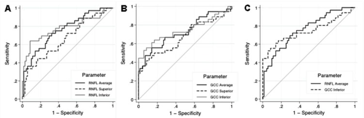
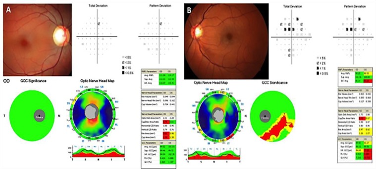
We evaluated the ability of spectral-domain optic coherence tomography (SD-OCT) to differentiate large physiological optic disc cupping (LPC) from glaucomatous cupping in eyes with intraocular pressure (IOP) within the normal range. We prospectively enrolled patients with glaucoma or presumed LPC. Participants had optic discs with confirmed or suspected glaucomatous damage (defined as a vertical cup-to-disc ratio≥0.6), and all eyes had known untreated IOP<21 mmHg. For glaucomatous eyes, a reproducible glaucomatous visual field (VF) defect was required. LPC eyes required normal VF and no evidence of progressive glaucomatous neuropathy (follow-up≥30 months). Peripapillary retinal nerve fiber layer (pRNFL) and macular ganglion cell complex (GCC) thicknesses were obtained using SD-OCT. For all studied parameters of pRNFL and GCC thicknesses, eyes with glaucoma (n=36) had significantly thinner values compared to eyes with LPC (n=71; P<0.05 for all comparisons). In addition, pRNFL parameters had sensitivity of 66.7% and specificity of 83.1%, and GCC parameters had sensitivity of 61.2% and specificity of 81.7%. The combination of the two analyses increased the sensitivity to 80.6%. In conclusion, while evaluating patients with large optic disc cupping and IOP in the statistically normal range, SD-OCT had only limited diagnostic ability to differentiate those with and without glaucoma. Although the diagnostic ability of the pRNFL and the GCC scans were similar, these parameters yielded an increase in sensitivity when combined, suggesting that both parameters could be considered simultaneously in these cases.
Keywords: Diagnostic Ability; Glaucoma; OCT; Optic Disc; SD-OCT.
Figures
References
-
- Shaffer R. Glaucoma suspect' or 'ocular hypertension? Arch Ophthalmol. 1977 Apr;95(4) PMID: 849180. - PubMed
-
- Kolker AE, Becker B. Ocular hypertension vs open-angle glaucoma: a different view. Arch Ophthalmol. 1977 Apr;95(4):586–7. PMID: 849179. - PubMed
-
- Phelps CD. Ocular hypertension: to treat or not to treat. Arch Ophthalmol. 1977 Apr;95(4):588–9. PMID: 849181. - PubMed
-
- Kitazawa Y, Horie T, Aoki S, Suzuki M, Nishioka K. Untreated ocular hypertension A long-term prospective study. Arch Ophthalmol. 1977 Jul;95(7):1180–4. PMID: 880076. - PubMed
-
- Wilensky JT, Podos SM, Becker B. Prognostic indicators in ocular hypertension. Arch Ophthalmol. 1974 Mar;91(3):200–2. PMID: 4814967. - PubMed
LinkOut - more resources
Full Text Sources