

Nanoparticles of copper stimulate angiogenesis at systemic and molecular level
- PMID: 25741768
- PMCID: PMC4394452
- DOI: 10.3390/ijms16034838
Nanoparticles of copper stimulate angiogenesis at systemic and molecular level
Abstract
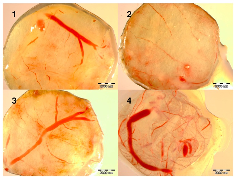
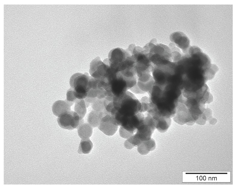
Copper is a key element affecting blood vessel growth and muscle development. However, the ions released from Cu salts are toxic. Given their specific physicochemical properties, nanoparticles of Cu (NanoCu) may have different bioactivity and affect the development of blood vessel and muscles in a different manner than Cu salts. The objective of the study was to evaluate the influence of NanoCu on embryo development and angiogenesis at the systemic and molecular level, in experiments using a chick embryo model. Fertilized chicken eggs were divided into a control group, and groups injected with a placebo, CuSO4 or NanoCu. Embryo development at the whole body level and molecular indices using an embryo chorioallantoic membrane model were measured during embryogenesis. The present study indicated for the first time that NanoCu have pro-angiogenic properties at the systemic level, to a greater degree than CuSO4 salt. The properties of NanoCu were confirmed at the molecular level, demonstrating significant effects on mRNA concentration and on mRNA gene expression of all pro-angiogenic and pro-proliferative genes measured herein.
Figures
Similar articles
-
In ovo administration of copper nanoparticles and copper sulfate positively influences chicken performance.J Sci Food Agric. 2016 Jul;96(9):3058-62. doi: 10.1002/jsfa.7477. Epub 2015 Oct 29. J Sci Food Agric. 2016. PMID: 26417698
-
Effect of copper nanoparticles on the mineral content of tissues and droppings, and growth of chickens.Arch Anim Nutr. 2018 Oct;72(5):396-406. doi: 10.1080/1745039X.2018.1505146. Epub 2018 Aug 13. Arch Anim Nutr. 2018. PMID: 30183391
-
In ovo leptin administration inhibits chorioallantoic membrane angiogenesis in female chicken embryos through the STAT3-mediated vascular endothelial growth factor (VEGF) pathway.Domest Anim Endocrinol. 2012 Jul;43(1):26-36. doi: 10.1016/j.domaniend.2012.01.007. Epub 2012 Mar 3. Domest Anim Endocrinol. 2012. PMID: 22417645
-
In ovo administration of human recombinant leptin shows dose dependent angiogenic effect on chicken chorioallantoic membrane.Biol Res. 2015 Jun 10;48(1):29. doi: 10.1186/s40659-015-0021-z. Biol Res. 2015. PMID: 26060038 Free PMC article.
-
Chick embryo chorioallantoic membrane as a useful tool to study angiogenesis.Int Rev Cell Mol Biol. 2008;270:181-224. doi: 10.1016/S1937-6448(08)01405-6. Int Rev Cell Mol Biol. 2008. PMID: 19081537 Review.
Cited by
-
Down-regulation of TGF-β, VEGF, and bFGF in vascular endothelial cells of chicken induced by a brittle star (Ophiocoma erinaceus) extract.Heliyon. 2020 Jan 16;6(1):e03199. doi: 10.1016/j.heliyon.2020.e03199. eCollection 2020 Jan. Heliyon. 2020. PMID: 31970303 Free PMC article.
-
Use of Metallic Nanoparticles Against Eimeria-the Coccidiosis-Causing Agents: A Comprehensive Review.Biol Trace Elem Res. 2025 Jun;203(6):3412-3431. doi: 10.1007/s12011-024-04399-8. Epub 2024 Oct 2. Biol Trace Elem Res. 2025. PMID: 39354182 Review.
-
Nanotechnology for angiogenesis: opportunities and challenges.Chem Soc Rev. 2020 Jul 21;49(14):5008-5057. doi: 10.1039/c8cs01021h. Epub 2020 Jun 15. Chem Soc Rev. 2020. PMID: 32538379 Free PMC article. Review.
-
Copper Preserves Vasculature Structure and Function by Protecting Endothelial Cells from Apoptosis in Ischemic Myocardium.J Cardiovasc Transl Res. 2021 Dec;14(6):1146-1155. doi: 10.1007/s12265-021-10128-6. Epub 2021 May 17. J Cardiovasc Transl Res. 2021. PMID: 33999373
-
Differential antimicrobial and cellular response of electrolytically metalized halloysite nanotubes having different amounts of surface metallization.Mater Adv. 2020 Sep 1;1(6):1705-1715. doi: 10.1039/d0ma00134a. Epub 2020 Jul 15. Mater Adv. 2020. PMID: 35813570 Free PMC article.
References
-
- Linder M.C., Goode C.A. Biochemistry of Copper. Springer–Verlag New York, LLC; New York, NY, USA: 1991. pp. 1–413.
-
- Shinkaruk S., Bayle M., Lain G., Deleris G. Vascular endothelial cell growth factor (VEGF), an emerging target for cancer chemotherapy. Curr. Med. Chem. 2003;3:95–117. - PubMed
Publication types
MeSH terms
Substances
LinkOut - more resources
Full Text Sources
Other Literature Sources
