. 2015 Mar 5;10(3):e0118287.
doi: 10.1371/journal.pone.0118287. eCollection 2015.

Sarsaparilla (Smilax Glabra Rhizome) extract inhibits migration and invasion of cancer cells by suppressing TGF-β1 pathway

Affiliations

Sarsaparilla (Smilax Glabra Rhizome) extract inhibits migration and invasion of cancer cells by suppressing TGF-β1 pathway

Tiantian She et al. PLoS One. .

Abstract

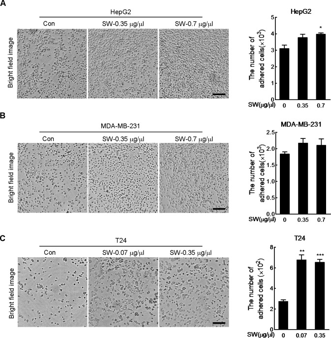
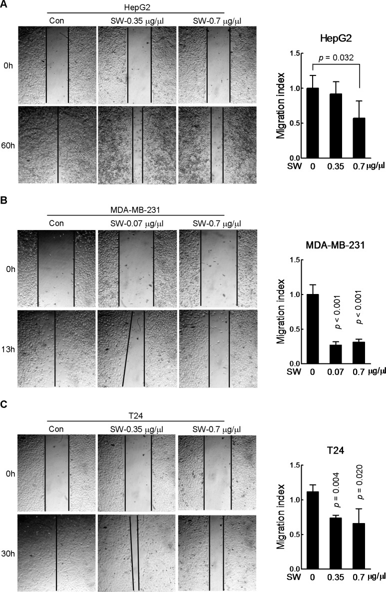
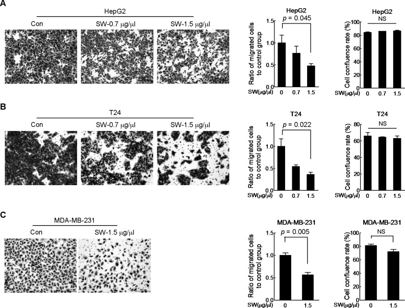
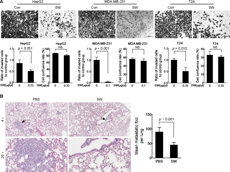
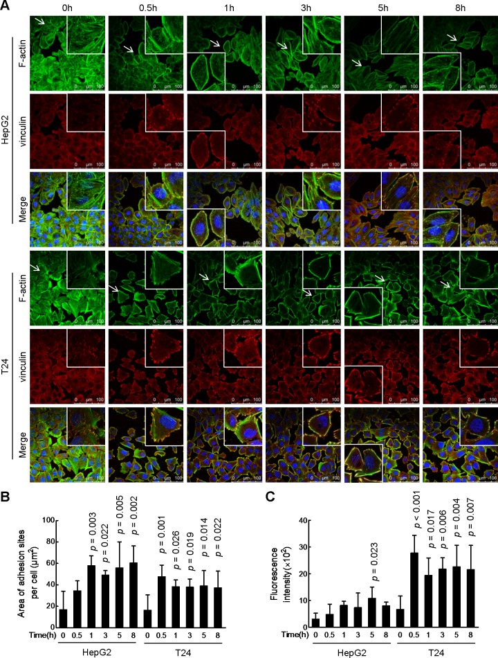
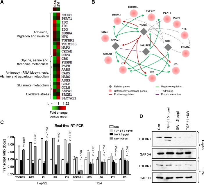
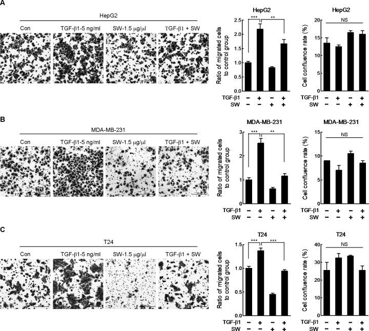
Sarsaparilla, also known as Smilax Glabra Rhizome (SGR), was shown to modulate immunity, protect against liver injury, lower blood glucose and suppress cancer. However, its effects on cancer cell adhesion, migration and invasion were unclear. In the present study, we found that the supernatant of water-soluble extract from SGR (SW) could promote adhesion, inhibit migration and invasion of HepG2, MDA-MB-231 and T24 cells in vitro, as well as suppress metastasis of MDA-MB-231 cells in vivo. Results of F-actin and vinculin dual staining showed the enhanced focal adhesion in SW-treated cells. Microarray analysis indicated a repression of TGF-β1 signaling by SW treatment, which was verified by real-time RT-PCR of TGF-β1-related genes and immunoblotting of TGFBR1 protein. SW was also shown to antagonize TGF-β1-promoted cell migration. Collectively, our study revealed a new antitumor function of Sarsaparilla in counteracting invasiveness of a subset of cancer cells by inhibiting TGF-β1 signaling.


Conflict of interest statement

Competing Interests: The authors have declared that no competing interests exist.

Figures

Fig 1
Fig 1. SW promotes cell adhesion.
Left panels: HepG2 (A), MDA-MB-231 (B) and T24 cells (C) were pretreated with indicated doses of SW for 15 or 30 min before seeding into the matrigel-coated wells, and 2 h later, adhered cells were counted after removing the floating cells by PBS. Representative images were displayed. Scale bar, 200 μm. Right panels: quantification of the data in the left panel, shown were composite results of three independently experiments with triplicate. Columns, mean; bars, SD.
Fig 2
Fig 2. SW inhibits cell scratch wound healing.
In vitro scratch assay was used to evaluate the effect of SW on the migration of HepG2 (A), MDA-MB-231 (B) and T24 cells (C). Representative images were displayed in left panel; quantification of the data in left panel was shown in right panel, shown were composite results of three independently experiments with triplicate parallel samples. The migration index represents migration speed in relative to control group. Columns, mean; bars, SD.
Fig 3
Fig 3. SW inhibits cell migration.
HepG2 (A), T24 (B) and MDA-MB-231 (C) cells were seeded into the transwell insert in the presence of indicated doses of SW for 12 h. Cells reaching the bottom side of the transwell membrane were stained and representative images were displayed in the upper panel. Scale bar, 200 μm. The data in left panel were quantified and shown in the middle panel, and the cell viability was reflected by cell confluence rate in the right panel. Shown were composite results of two independently experiments. Columns, mean; bars, SD; NS, not statistically significant.
Fig 4
Fig 4. SW inhibits cell invasion in vitro and metastasis in vivo.
(A) The transwell chambers were pre-coated with 60 μl matrigel dilution (1:4 in serum-free medium) before seeding cells. Cells were incubated with indicated doses of SW for 36 h before staining. Representative images were displayed in the upper panel. Scale bar, 200 μm. The data of migration and cell viability were quantified and shown respectively in lower panel, shown were composite results of two independently experiments with triplicate. Columns, mean; bars, SD; NS, not statistically significant. (B) Left panel: representative pictures of H&E stained lung tissues in PBS- and SW-treated group. Paraffin-embedded lungs were sectioned at 30-μm intervals and > 10 MDA-MB-231 cancer cells were identified as metastatic foci. Magnification, 4× (upper) and 20× (lower). Right panel: quantification of lung metastatic foci in PBS- and SW-treated mice group (n = 8 for each group). Columns, mean; bars, SD.
Fig 5
Fig 5. SW increases size and fluorescence intensity of focal adhesion.
(A) HepG2 and T24 cells were treated with SW (3.5 μg/μl) for indicated times and then immuno-stained with F-actin (FITC-phalloidin) and vinculin (red). Nuclei were counterstained by DAPI (blue). Representative images were displayed. Scale bar, 100 μm. (B) The area of adhesion sites per cell (where F-actin and vinculin merged). Shown were composite results of two independently experiments (n = 50 cells). Columns, mean; bars, SD. (C) The fluorescence intensity of vinculin in the adhesion patches per cell. Shown were composite results of two independently experiments (n = 50 cells). Columns, mean; bars, SD.
Fig 6
Fig 6. Microarray analysis result indicated repression of TGF-β1 pathway in SW-treated cells.
(A) The heat map of functional enrichment of differentialy expressed genes from the HepG2 chip result. Cluster 3.0 and TreeView software were used for generating the heat map. Red: up-regulation versus mean; green: down-regulation versus mean. (B) The regulatory network among the genes related to adhesion, migration and invasion in (A) using the STRING database. (C) Real-time RT-PCR verification of the genes in HepG2 and T24 cells treated with TGF-β1 with or without SW. Shown were composite results of three independently experiments with triplicate. Columns, mean; bars, SD. (D) Effect of SW on TGF-β1-induced up-regulation of TGFBR1 was evaluated by immunoblotting. Shown were representative results from 3 independent experiments.
Fig 7
Fig 7. SW abrogates TGF-β1-induced boost in migration.
HepG2 (A), MDA-MB-231 (B) and T24 (C) cells were seeded into the transwell inserts in the presence of TGF-β1 plus SW. 12 h later, Cells were stained and pictured. Representative images were displayed in the left panel. Scale bar, 100 μm. The data of migration were quantified and shown in the middle panel. The cell viability was reflected by cell confluence rate in the right panel. Shown was composite results of two independently experiments with triplicate. Columns, mean; bars, SD; NS, not statistically significant.

References

    1. Tse TW (2003) Use of common Chinese herbs in the treatment of psoriasis. Clin Exp Dermatol. 28: 469–475. - PubMed
    1. Ulbricht C, Basch E (2001) Natural Standard. Database: Foods, Herbs & Supplements [Internet]. Available: http://www.naturalstandard.com/databases/herbssupplements/all/a/.
    1. Chen L, Yin Y, Yi H, Xu Q, Chen T (2007) Simultaneous quantification of five major bioactive flavonoids in Rhizoma smilacis glabrae by high-performance liquid chromatography. J Pharm Biomed Anal. 43: 1715–1720. - PubMed
    1. Shi L, Xu L, Yang Y, Song H, Pan H, et al. (2013) Suppressive effect of modified Simiaowan on experimental gouty arthritis: an in vivo and in vitro study. J Ethnopharmacol. 150: 1038–1044. 10.1016/j.jep.2013.10.023 - DOI - PubMed
    1. Jiang J, Xu Q (2003) Immunomodulatory activity of the aqueous extract from rhizome of Smilax glabra in the later phase of adjuvant-induced arthritis in rats. J Ethnopharmacol. 85: 53–59. - PubMed

Publication types

Substances

Associated data