. 2015 Mar 5;10(3):e0119899.
doi: 10.1371/journal.pone.0119899. eCollection 2015.

Conformational dynamics and antigenicity in the disordered malaria antigen merozoite surface protein 2

Affiliations

Conformational dynamics and antigenicity in the disordered malaria antigen merozoite surface protein 2

Christopher A MacRaild et al. PLoS One. .

Abstract

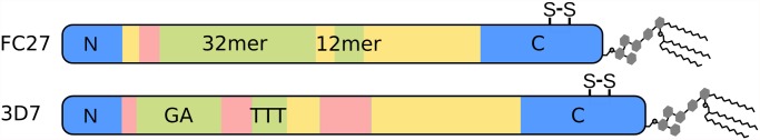
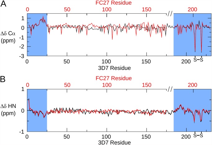
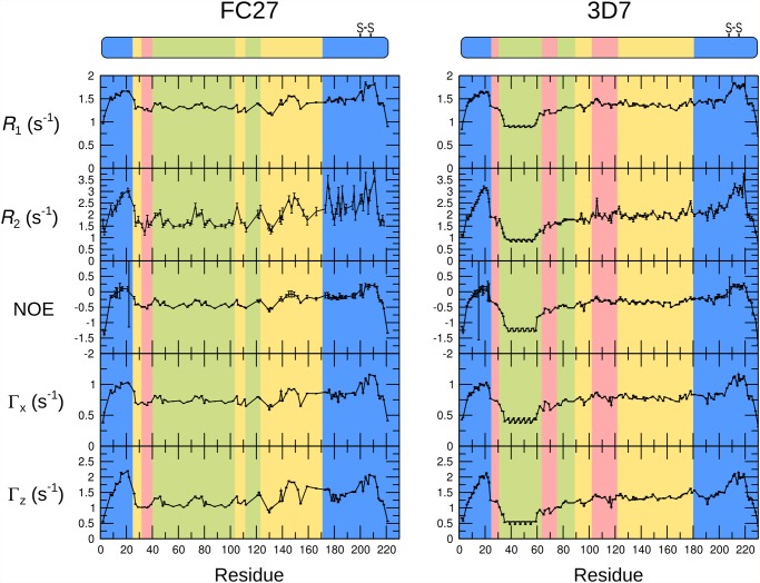
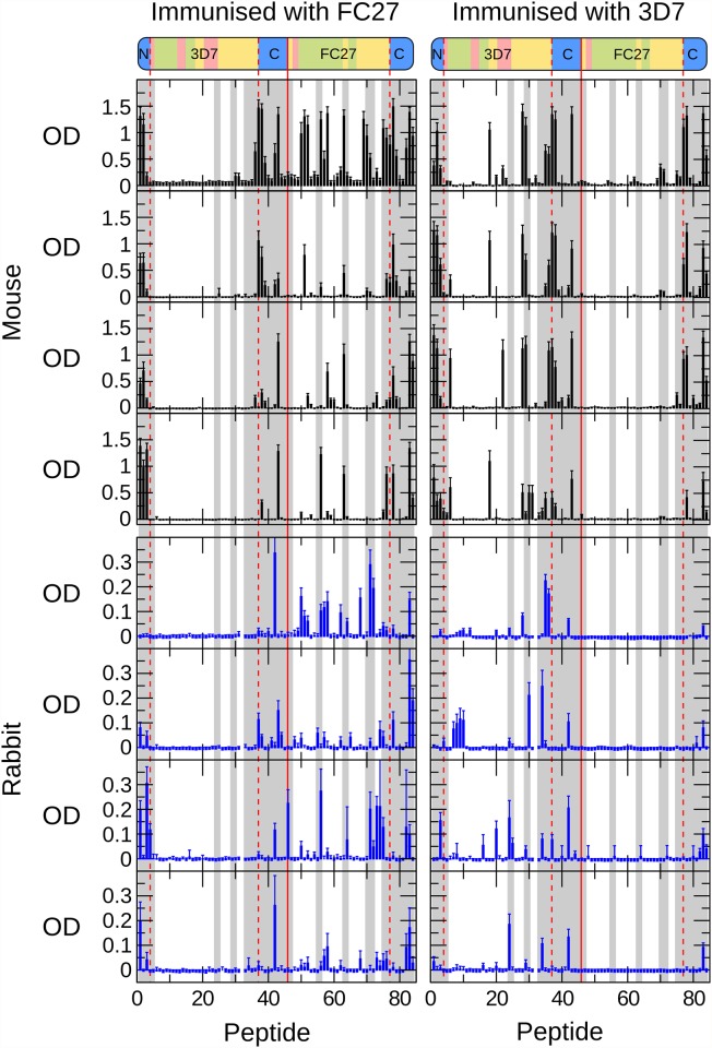
Merozoite surface protein 2 (MSP2) of Plasmodium falciparum is an abundant, intrinsically disordered protein that is GPI-anchored to the surface of the invasive blood stage of the malaria parasite. Recombinant MSP2 has been trialled as a component of a malaria vaccine, and is one of several disordered proteins that are candidates for inclusion in vaccines for malaria and other diseases. Nonetheless, little is known about the implications of protein disorder for the development of an effective antibody response. We have therefore undertaken a detailed analysis of the conformational dynamics of the two allelic forms of MSP2 (3D7 and FC27) using NMR spectroscopy. Chemical shifts and NMR relaxation data indicate that conformational and dynamic properties of the N- and C-terminal conserved regions in the two forms of MSP2 are essentially identical, but significant variation exists between and within the central variable regions. We observe a strong relationship between the conformational dynamics and the antigenicity of MSP2, as assessed with antisera to recombinant MSP2. Regions of increased conformational order in MSP2, including those in the conserved regions, are more strongly antigenic, while the most flexible regions are minimally antigenic. This suggests that modifications that increase conformational order may offer a means to tune the antigenicity of MSP2 and other disordered antigens, with implications for vaccine design.


Conflict of interest statement

Competing Interests: James Beeson is a PLOS ONE Editorial Board member. This does not alter the authors' adherence to PLOS ONE Editorial policies and criteria.

Figures

Fig 1
Fig 1. Schematic depiction of the primary structure of the two allelic families of MSP2.
The conserved N- and C-terminal regions of MSP2 are in blue, while the allele-specific central region is composed of polymorphic repeats (green) and non-repetitive sequences (pink) as well as dimorphic regions (yellow) that differ between the allelic families but are conserved within them. The position of the conserved disulfide bond in the C-terminal regions is indicated.
Fig 2
Fig 2. 1H,15N heteronuclear single-quantum correlation spectra of 3D7 (blue) and FC27 (red) MSP2.
Assigned peaks are labelled in the expanded regions, highlighting the similarity of chemical shift for these residues in the conserved N- and C-terminal regions (labelled in black type, FC27 numbering) of the two allelic forms of MSP2.
Fig 3
Fig 3. Secondary chemical shifts of MSP2.
The difference between the observed Cα (A) and HN (B) chemical shifts and those predicted for a disordered protein by the method of Tamiola et al. [50] is plotted for 3D7 (black) and FC27 (red) MSP2. The data for FC27 are plotted on a broken axis (top) in order to correctly align the conserved regions.
Fig 4
Fig 4. The 15N auto-relaxation rates R 1, R 2, cross-correlated relaxation rates Γx, Γz, and steady-state 15N-1H NOE for FC27 (left) and 3D7 (right) MSP2.
The sequence regions of MSP2 are colour-coded as in Fig. 1.
Fig 5
Fig 5. Values of the 15N spectral density functions for FC27 (left) and 3D7 (right) MSP2, as determined from reduced spectral density mapping from the auto-relaxation rates and steady-state 15N-1H NOE (black) and cross-correlated relaxation rates (red).
The sequence regions of MSP2 are colour-coded as in Fig. 1.
Fig 6
Fig 6. Antigenic profile of MSP2 mapped by ELISA.
Four mice (top panels, black bars) and four rabbits (bottom panels, blue bars) were immunised with either FC27 (left panels) or 3D7 (right panels) MSP2. Individual immune sera were tested against a single panel of overlapping peptides covering the sequences of both 3D7 MSP2 (peptides 1–45) and FC27 MSP2 (peptides 46–84), as shown schematically above. The conserved N terminal (peptides 1–3) and C terminal (peptides 37–45 and 77–84) regions are common to both 3D7 and FC27 MSP2 and are delineated with dashed red lines. Peptides showing greater than the median level of conformational restriction are shaded grey (Table 1). Mean optical density from triplicate assays is plotted for each serum, corrected for the response from unimmunised control sera. Error bars are one standard deviation.
Fig 7
Fig 7. Comparison of experimental patterns of antigenicity, predicted antigenicity, and conformational dynamics, for FC27 (left) and 3D7 (right) MSP2.
A. Antigenicity profiles of MSP2 inferred from experimental immunisation of mice (black) and rabbits (red) are plotted against the sequence. Black bars (top) denote the location of epitopes of a panel of monoclonal antibodies to MSP2. B. Conformational flexibility of MSP2 as measured by the spectral density functions derived from the 15N relaxation data. Spectral density functions are plotted at zero frequency (black line) and at the 15N Larmor frequency (red line). C. Antigenicity of MSP2 as predicated using BepiPred [60] (red, right axis) and the method of Kolaskar and Tongaonkar [61] (black, left axis). The threshold for epitope prediction for both methods is denoted by the grey line.

Similar articles

Cited by

References

    1. Uversky VN, Oldfield CJ, Dunker AK. Showing your ID: intrinsic disorder as an ID for recognition, regulation and cell signaling. 2005; J Mol Recognit 18: 343–384. - PubMed
    1. Oldfield CJ, Dunker AK. Intrinsically disordered proteins and intrinsically disordered protein regions. 2014; Annu Rev Biochem 83: 553–584. 10.1146/annurev-biochem-072711-164947 - DOI - PubMed
    1. Xue B, Blocquel D, Habchi J, Uversky AV, Kurgan L, Uversky VN, et al. Structural disorder in viral proteins. 2014; Chem Rev 114: 6880–6911. 10.1021/cr4005692 - DOI - PubMed
    1. Feng ZP, Zhang X, Han P, Arora N, Anders RF, Norton RS. Abundance of intrinsically unstructured proteins in P. falciparum and other apicomplexan parasite proteomes. 2006; Mol Biochem Parasitol 150: 256–267. - PubMed
    1. Pavlovic MD, Jandrlic DR, Mitic NS. Epitope distribution in ordered and disordered protein regions. Part B—Ordered regions and disordered binding sites are targets of T- and B-cell immunity. 2014; J Immunol Methods 407: 90–107. 10.1016/j.jim.2014.03.027 - DOI - PubMed

Publication types

MeSH terms

Substances