

miR-186 and 326 predict the prognosis of pancreatic ductal adenocarcinoma and affect the proliferation and migration of cancer cells
- PMID: 25742499
- PMCID: PMC4351009
- DOI: 10.1371/journal.pone.0118814
miR-186 and 326 predict the prognosis of pancreatic ductal adenocarcinoma and affect the proliferation and migration of cancer cells
Abstract
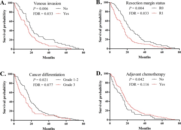
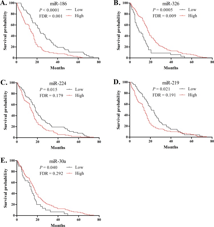
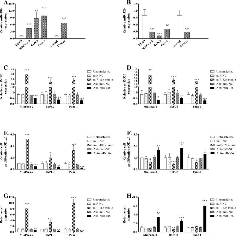
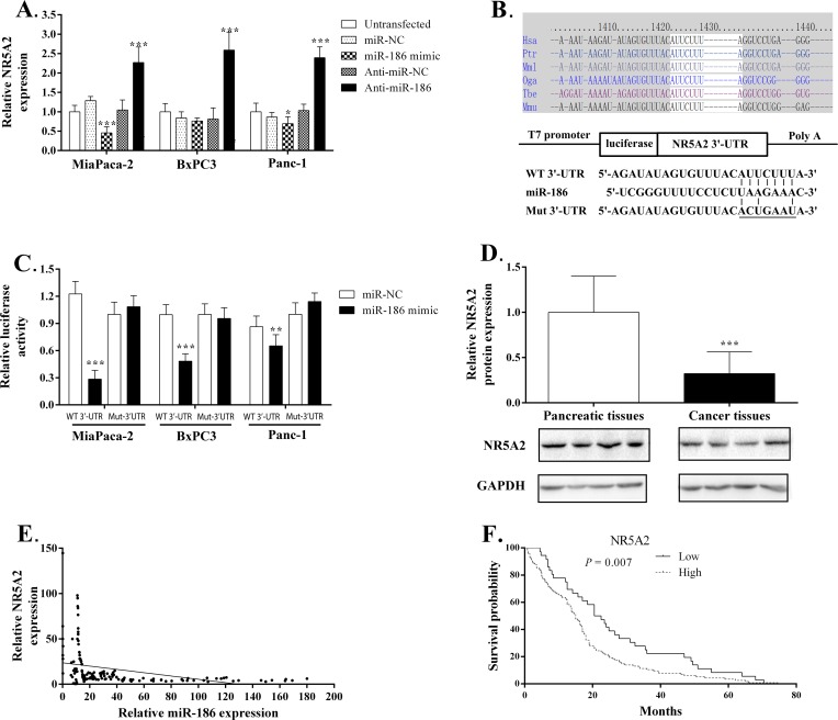
MicroRNAs can function as key tumor suppressors or oncogenes and act as biomarkers for cancer diagnosis or prognosis. Although high-throughput assays have revealed many miRNA biomarkers for pancreatic ductal adenocarcinoma (PDAC), only a few have been validated in independent populations or investigated for functional significance in PDAC pathogenesis. In this study, we correlated the expression of 36 potentially prognostic miRNAs within PDAC tissue with clinico-pathological features and survival in 151 Chinese patients. We then analyzed the functional roles and target genes of two miRNAs in PDAC development. We found that high expression of miR-186 and miR-326 predict poor and improved survival, respectively. miR-186 was over-expressed in PDAC patients compared with controls, especially in patients with large tumors (>2 cm), lymph node metastasis, or short-term survival (< 24 months). In contrast, miR-326 was down-regulated in patients compared with controls and displayed relatively increased expression in the patients with long-term survival or without venous invasion. Functional experiments revealed that PDAC cell proliferation and migration was decreased following inhibition and enhanced following over-expression of miR-186. In contrast, it was enhanced following inhibition and decreased after over-expression of miR-326. A luciferase assay indicated that miR-186 can bind directly to the 3'-UTR of NR5A2 to repress gene expression. These findings suggest that miR-186 over-expression contributes to the invasive potential of PDAC, likely via suppression of NR5A2, thereby leading to a poor prognosis; high miR-326 expression prolongs survival likely via the decreasing invasive potential of PDAC cells. These two miRNAs can be used as markers for clinical diagnosis and prognosis, and they represent therapeutic targets for PDAC.
Conflict of interest statement
Figures
Similar articles
-
MicroRNA-183 is involved in cell proliferation, survival and poor prognosis in pancreatic ductal adenocarcinoma by regulating Bmi-1.Oncol Rep. 2014 Oct;32(4):1734-40. doi: 10.3892/or.2014.3374. Epub 2014 Aug 1. Oncol Rep. 2014. PMID: 25109303
-
MicroRNA-323-3p inhibits cell invasion and metastasis in pancreatic ductal adenocarcinoma via direct suppression of SMAD2 and SMAD3.Oncotarget. 2016 Mar 22;7(12):14912-24. doi: 10.18632/oncotarget.7482. Oncotarget. 2016. PMID: 26908446 Free PMC article.
-
Downregulated miR-98-5p promotes PDAC proliferation and metastasis by reversely regulating MAP4K4.J Exp Clin Cancer Res. 2018 Jul 3;37(1):130. doi: 10.1186/s13046-018-0807-2. J Exp Clin Cancer Res. 2018. PMID: 29970191 Free PMC article.
-
Clinical potential of microRNAs in pancreatic ductal adenocarcinoma.Pancreas. 2011 Nov;40(8):1165-71. doi: 10.1097/MPA.0b013e3182218ffb. Pancreas. 2011. PMID: 22001830 Review.
-
MicroRNA in pancreatic adenocarcinoma: predictive/prognostic biomarkers or therapeutic targets?Oncotarget. 2015 Sep 15;6(27):23323-41. doi: 10.18632/oncotarget.4492. Oncotarget. 2015. PMID: 26259238 Free PMC article. Review.
Cited by
-
The Dual Role of miR-186 in Cancers: Oncomir Battling With Tumor Suppressor miRNA.Front Oncol. 2020 Mar 5;10:233. doi: 10.3389/fonc.2020.00233. eCollection 2020. Front Oncol. 2020. PMID: 32195180 Free PMC article. Review.
-
MicroRNA-326 suppresses the proliferation, migration and invasion of cervical cancer cells by targeting ELK1.Oncol Lett. 2017 May;13(5):2949-2956. doi: 10.3892/ol.2017.5852. Epub 2017 Mar 13. Oncol Lett. 2017. PMID: 28529556 Free PMC article.
-
TBL1XR1 promotes migration and invasion in osteosarcoma cells and is negatively regulated by miR-186-5p.Am J Cancer Res. 2018 Dec 1;8(12):2481-2493. eCollection 2018. Am J Cancer Res. 2018. PMID: 30662805 Free PMC article.
-
MicroRNA-186 regulates the invasion and metastasis of bladder cancer via vascular endothelial growth factor C.Exp Ther Med. 2017 Oct;14(4):3253-3258. doi: 10.3892/etm.2017.4908. Epub 2017 Aug 8. Exp Ther Med. 2017. PMID: 28966690 Free PMC article.
-
The Emerging Role of miRNAs for the Radiation Treatment of Pancreatic Cancer.Cancers (Basel). 2020 Dec 9;12(12):3703. doi: 10.3390/cancers12123703. Cancers (Basel). 2020. PMID: 33317198 Free PMC article.
References
-
- Saif MW (2006) Pancreatic cancer: highlights from the 42nd annual meeting of the American Society of Clinical Oncology, 2006. JOP 7: 337–348. - PubMed
-
- Hezel AF, Kimmelman AC, Stanger BZ, Bardeesy N, Depinho RA (2006) Genetics and biology of pancreatic ductal adenocarcinoma. Genes Dev 20: 1218–1249. - PubMed
Publication types
MeSH terms
Substances
LinkOut - more resources
Full Text Sources
Other Literature Sources
Medical
