

Boon and Bane of Inflammation in Bone Tissue Regeneration and Its Link with Angiogenesis
- PMID: 25742724
- PMCID: PMC4533093
- DOI: 10.1089/ten.TEB.2014.0677
Boon and Bane of Inflammation in Bone Tissue Regeneration and Its Link with Angiogenesis
Abstract
Delayed healing or nonhealing of bone is an important clinical concern. Although bone, one of the two tissues with scar-free healing capacity, heals in most cases, healing is delayed in more than 10% of clinical cases. Treatment of such delayed healing condition is often painful, risky, time consuming, and expensive. Tissue healing is a multistage regenerative process involving complex and well-orchestrated steps, which are initiated in response to injury. At best, these steps lead to scar-free tissue formation. At the onset of healing, during the inflammatory phase, stationary and attracted macrophages and other immune cells at the fracture site release cytokines in response to injury. This initial reaction to injury is followed by the recruitment, proliferation, and differentiation of mesenchymal stromal cells, synthesis of extracellular matrix proteins, angiogenesis, and finally tissue remodeling. Failure to heal is often associated with poor revascularization. Since blood vessels mediate the transport of circulating cells, oxygen, nutrients, and waste products, they appear essential for successful healing. The strategy of endogenous regeneration in a tissue such as bone is interesting to analyze since it may represent a blueprint of successful tissue formation. This review highlights the interdependency of the time cascades of inflammation, angiogenesis, and tissue regeneration. A better understanding of these inter-relations is mandatory to early identify patients at risk as well as to overcome critical clinical conditions that limit healing. Instead of purely tolerating the inflammatory phase, modulations of inflammation (immunomodulation) might represent a valid therapeutic strategy to enhance angiogenesis and foster later phases of tissue regeneration.
Figures

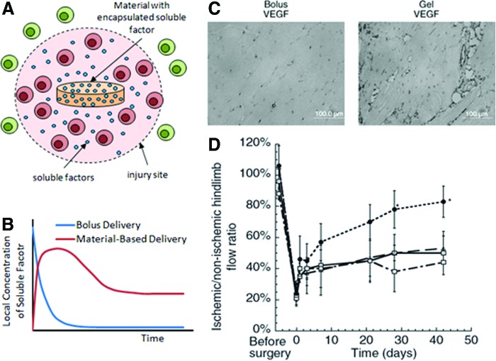
 ), and material delivery of VEGF (●). Images adopted from Silva and Mooney. Color images available online at
 Similar articles
-
Initial immune reaction and angiogenesis in bone healing.J Tissue Eng Regen Med. 2014 Feb;8(2):120-30. doi: 10.1002/term.1505. Epub 2012 Apr 11. J Tissue Eng Regen Med. 2014. PMID: 22495762
-
Bringing new life to damaged bone: the importance of angiogenesis in bone repair and regeneration.Bone. 2015 Jan;70:19-27. doi: 10.1016/j.bone.2014.09.017. Epub 2014 Sep 28. Bone. 2015. PMID: 25263520 Review.
-
CD31+ Cells From Peripheral Blood Facilitate Bone Regeneration in Biologically Impaired Conditions Through Combined Effects on Immunomodulation and Angiogenesis.J Bone Miner Res. 2017 May;32(5):902-912. doi: 10.1002/jbmr.3062. Epub 2017 Mar 1. J Bone Miner Res. 2017. PMID: 27976803
-
Inflammatory phase of bone healing initiates the regenerative healing cascade.Cell Tissue Res. 2012 Mar;347(3):567-73. doi: 10.1007/s00441-011-1205-7. Epub 2011 Jul 26. Cell Tissue Res. 2012. PMID: 21789579
-
The decisive early phase of bone regeneration.Nat Rev Rheumatol. 2023 Feb;19(2):78-95. doi: 10.1038/s41584-022-00887-0. Epub 2023 Jan 9. Nat Rev Rheumatol. 2023. PMID: 36624263 Review.
Cited by
-
Complex Spatio-Temporal Interplay of Distinct Immune and Bone Cell Subsets during Bone Fracture Healing.Cells. 2023 Dec 24;13(1):40. doi: 10.3390/cells13010040. Cells. 2023. PMID: 38201244 Free PMC article.
-
Clinical pathologies of bone fracture modelled in zebrafish.Dis Model Mech. 2019 Sep 3;12(9):dmm037630. doi: 10.1242/dmm.037630. Dis Model Mech. 2019. PMID: 31383797 Free PMC article.
-
The Role of Sex Differences in Bone Health and Healing.Biology (Basel). 2023 Jul 12;12(7):993. doi: 10.3390/biology12070993. Biology (Basel). 2023. PMID: 37508423 Free PMC article. Review.
-
Immune Modulation to Enhance Bone Healing-A New Concept to Induce Bone Using Prostacyclin to Locally Modulate Immunity.Front Immunol. 2019 Apr 5;10:713. doi: 10.3389/fimmu.2019.00713. eCollection 2019. Front Immunol. 2019. PMID: 31024548 Free PMC article.
-
Mesoporous silicate nanoparticles/3D nanofibrous scaffold-mediated dual-drug delivery for bone tissue engineering.J Control Release. 2018 Jun 10;279:69-78. doi: 10.1016/j.jconrel.2018.04.011. Epub 2018 Apr 9. J Control Release. 2018. PMID: 29649529 Free PMC article.
References
-
- Kolar P., Schmidt-Bleek K., Schell H., Gaber T., Toben D., Schmidmaier G., Perka C., Buttgereit F., and Duda G.N. The early fracture hematoma and its potential role in fracture healing. Tissue Eng Part B Rev 16, 427, 2010 - PubMed
-
- Einhorn T.A., Majeska R.J., Rush E.B., Levine P.M., and Horowitz M.C. The expression of cytokine activity by fracture callus. J Bone Miner Res 10, 1272, 1995 - PubMed
-
- Kon T., Cho T.J., Aizawa T., Yamazaki M., Nooh N., Graves D., Gerstenfeld L.C., and Einhorn T.A. Expression of osteoprotegerin, receptor activator of NF-kappaB ligand (osteoprotegerin ligand) and related proinflammatory cytokines during fracture healing. J Bone Miner Res 16, 1004, 2001 - PubMed
-
- Schmidt-Bleek K., Petersen A., Dienelt A., Schwarz C., and Duda G.N. Initiation and early control of tissue regeneration—bone healing as a model system for tissue regeneration. Expert Opin Biol Ther 14, 247, 2014 - PubMed
Publication types
MeSH terms
Grants and funding
LinkOut - more resources
Full Text Sources
Other Literature Sources
