Review
. 2015 Jul:114:58-71.
doi: 10.1016/j.biochi.2015.02.017. Epub 2015 Mar 2.

Quantitative studies of mRNA recruitment to the eukaryotic ribosome

Affiliations
Review

Quantitative studies of mRNA recruitment to the eukaryotic ribosome

Christopher S Fraser. Biochimie. 2015 Jul.

Abstract

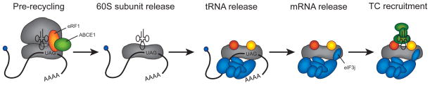
The process of peptide bond synthesis by ribosomes is conserved between species, but the initiation step differs greatly between the three kingdoms of life. This is illustrated by the evolution of roughly an order of magnitude more initiation factor mass found in humans compared with bacteria. Eukaryotic initiation of translation is comprised of a number of sub-steps: (i) recruitment of an mRNA and initiator methionyl-tRNA to the 40S ribosomal subunit; (ii) migration of the 40S subunit along the 5' UTR to locate the initiation codon; and (iii) recruitment of the 60S subunit to form the 80S initiation complex. Although the mechanism and regulation of initiation has been studied for decades, many aspects of the pathway remain unclear. In this review, I will focus discussion on what is known about the mechanism of mRNA selection and its recruitment to the 40S subunit. I will summarize how the 43S preinitiation complex (PIC) is formed and stabilized by interactions between its components. I will discuss what is known about the mechanism of mRNA selection by the eukaryotic initiation factor 4F (eIF4F) complex and how the selected mRNA is recruited to the 43S PIC. The regulation of this process by secondary structure located in the 5' UTR of an mRNA will also be discussed. Finally, I present a possible kinetic model with which to explain the process of mRNA selection and recruitment to the eukaryotic ribosome.

Keywords: 43S PIC; eIF4F; mRNA recruitment.


Figures

Figure 1
Figure 1
Pathway of eukaryotic translation initiation. In the first step of initiation (step 1), a mRNA is recruited to the 40S subunit. The eIF4F complex (eIF4E, eIF4A, and eIF4G) specifically binds to the m7G cap structure located on the 5′ end of the mRNA. Although not shown, an interaction between PABP, the poly(A) tail and eIF4F can also occur. The eIF4F bound mRNA is recruited to the 43S PIC by virtue of numerous interactions between initiation components described in the text. In the second step of initiation (step 2), the 40S subunit scans along the 5′ UTR in a 5′ to 3′ direction. Codons are continuously sampled by the initiator tRNA anticodon until one is selected (shown here as an AUG). In the final step of initiation (step 3), two GTP hydrolysis steps occur, resulting in the recruitment of the 60S subunit and initiation factor release. The newly formed 80S ribosome then enters the elongation cycle.
Figure 2
Figure 2
40S subunit structure and substrate binding sites. The atomic model of the yeast 40S subunit is shown from the interface view according to a recent 40S–mRNA–eIF1–eIF1A–TC cryo-EM model (PDB: 3J81 [36]). Landmarks for the 40S subunit are labeled: A, A-site; P, P-site; E, E-site; bk, beak; b, body; pt, platform; and h, head. The latch of the 40S is formed between helix 18 of the body and helix 34 of the head. The mRNA entry and exit channels either side of the neck are labeled accordingly. Binding sites of mRNA (40S–mRNA), eIF1 and eIF1A (40S–eIF1–eIF1A), TC (40S–eIF1–eIF1A–TC) are shown individually for clarity by removal of individual substrates from the original atomic model (PDB: 3J81). The general binding site for the bulk of the eIF3 complex is shown on the solvent exposed surface of the 40S subunit according to a recent low-resolution cryo-EM model of the mammalian 43S complex [38].
Figure 3
Figure 3
Ribosome recycling pathway in eukaryotes. One possible pathway of 43S PIC generation is shown according to recent models [63-65]. A termination/prerecycling complex containing eRF1 and ABCE1 is shown as the first complex in the pathway, although it should be noted that additional steps in the termination pathway exist prior to this step [17]. The ATPase activity of ABCE1 is responsible for the dissociation of the 60S subunit and eRF1 in the first step of the pathway. The deacylated tRNA in the P-site is then released, most likely upon recruitment of eIF1, eIF1A and eIF3. The mRNA is then released from the complex upon binding of eIF3j into the mRNA entry channel and A-site of the 40S subunit. Finally, the TC is recruited to the 40S subunit to form the 43S PIC. In addition to the labelling of eRF1 and ABCE1, individual initiation components are shown according to the key provided in Figure 1.
Figure 4
Figure 4
High-resolution structures of eIF4F components. (A) Cartoon representations of human eIF4GI and eIF4AI proteins. Colored boxes represent the general positions of conserved domains in each protein and are labelled accordingly. The binding sites of other initiation components with each conserved domain in eIF4G are indicated. (B) A selection of high-resolution structures for domains of eIF4G together with eIF4E and eIF4A are shown. Each structural model is colored according to the cartoons shown in panel A. The HEAT-1 domain of yeast eIF4G bound to eIF4AI is shown with eIF4AI in an “open” conformation (bound to AMP), which is not compatible with RNA binding (PDB: 2VSO [178]). As a comparison, the “closed” conformation of eIF4AIII from the exon junction complex is presented (bound to ADPNP), which is bound to RNA (PDB: 2HYI [179]). Other components of the exon-junction complex have been omitted for clarity. A solution structure of the yeast eIF4E bound to m7GDP (cap) and its eIF4G-binding domain is shown with eIF4E colored orange (PDB: 1RF8 [180]). A structure of the C-terminal HEAT-2 and HEAT-3 domains of human eIF4GI is shown in the absence of its eIF4AI and MNK1 binding partners (PDB: 1UG3 [181]). The N-terminal PAM domain of eIF4G is shown bound to RRM1 and RRM2 of PABP together with a short stretch of poly(A) mRNA (PDB: 4F02 [182]).
Figure 5
Figure 5
A possible kinetic pathway of eukaryotic mRNA recruitment. In the step 1 of the pathway, initial binding of the eIF4F complex to the m7G cap structure of the mRNA occurs through the eIF4E subunit of eIF4F. In step 2, any m7G cap proximal secondary structure must be melted to enable eIF4F accommodation to occur. This likely involves the interaction of eIF4G RNA binding domains with single stranded mRNA. In step 3, the eIF4F–mRNA complex is recruited to the 43S PIC. This step involves the positioning of single stranded mRNA into the decoding site of the 40S subunit. In step 4, additional mRNA secondary structure downstream of the m7G cap structure is melted as the 43S PIC migrates along the 5′ UTR in search of the initiation codon. While dissociation of the m7G cap structure from eIF4E is depicted during this step, the precise timing of eIF4E detachment from the m7G cap and/or eIF4G is not known. For each step in the pathway, forward and reverse rate constants are shown, as described in the text. Individual initiation components are shown according to the key provided in Figure 1. Although not shown, it should be noted that ATP hydrolysis is likely to accelerate each forward step.

Similar articles

Cited by

References

    1. Chu D, Kazana E, Bellanger N, Singh T, Tuite MF, von der Haar T. Translation elongation can control translation initiation on eukaryotic mRNAs. Embo J. 2014;33:21–34. - PMC - PubMed
    1. Plotkin JB, Kudla G. Synonymous but not the same: the causes and consequences of codon bias. Nature reviews Genetics. 2011;12:32–42. - PMC - PubMed
    1. Ciandrini L, Stansfield I, Romano MC. Ribosome traffic on mRNAs maps to gene ontology: genome-wide quantification of translation initiation rates and polysome size regulation. PLoS computational biology. 2013;9:e1002866. - PMC - PubMed
    1. Kemp AJ, Betney R, Ciandrini L, Schwenger AC, Romano MC, Stansfield I. A yeast tRNA mutant that causes pseudohyphal growth exhibits reduced rates of CAG codon translation. Molecular microbiology. 2013;87:284–300. - PMC - PubMed
    1. Zhou M, Guo J, Cha J, Chae M, Chen S, Barral JM, Sachs MS, Liu Y. Non-optimal codon usage affects expression, structure and function of clock protein FRQ. Nature. 2013;495:111–115. - PMC - PubMed

Publication types