

Transforming growth factor beta 3 involved in the pathogenesis of synovial chondromatosis of temporomandibular joint
- PMID: 25742744
- PMCID: PMC4351526
- DOI: 10.1038/srep08843
Transforming growth factor beta 3 involved in the pathogenesis of synovial chondromatosis of temporomandibular joint
Abstract
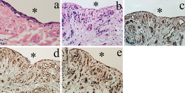
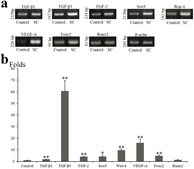
Synovial chondromatosis (SC) of temporomandibular joint is rare proliferative disorder featured by the formation of cartilaginous nodules in synovium and joint space. Transforming growth factor beta 3 (TGF-β3) is closely related to chondrogenic differentiation, and might participate in pathogenesis of SC. We discovered that increased quantity of synoviocytes and blood vessels were observed in SC synovium. The vessel wall and sublining fibroblasts were stained positively by the antibodies against TGF-β3, fibroblast growth factor 2 (FGF-2), and CD34. In loose bodies (LBs), TGF-β3 was mainly expressed in chondrocytes and FGF-2 was expressed in chondrocytes, fibroblasts, and vessel walls. Expressions of TGF-β1, TGF-β3, FGF-2, Sox9, Wnt-4, Foxc2, and VEGF-A mRNA were significantly higher in SC synovium. Stimulation of TGF-β3 on synoviocytes increased alkaline phosphatase (ALP) activity and expressions of chondrogenic genes (Sox9, Col2α1, Aggrecan, Wnt-4, and Wnt-11), osteogenic genes (Runx2, Foxc2, osteocalcin, and Col1α1), and VEGF-A, but failed to influence FGF-2 expression. However, the addition of FGF-2 increased TGF-β3 expression. In conclusion, TGF-β3 existed in synovium and LBs of SC, and was responsible for the pathogenesis of SC.
Figures


Similar articles
-
Fibroblast growth factor 2 involved in the pathogenesis of synovial chondromatosis of temporomandibular joint.J Oral Pathol Med. 2014 May;43(5):388-94. doi: 10.1111/jop.12146. Epub 2013 Dec 26. J Oral Pathol Med. 2014. PMID: 24372705
-
Role of synovium-derived fibrous cartilage in temporomandibular joint synovial chondromatosis.J Oral Pathol Med. 2019 Jan;48(1):79-86. doi: 10.1111/jop.12788. Epub 2018 Oct 26. J Oral Pathol Med. 2019. PMID: 30290017
-
CD34+ synovial fibroblasts exhibit high osteogenic potential in synovial chondromatosis.Cell Tissue Res. 2024 Jul;397(1):37-50. doi: 10.1007/s00441-024-03892-9. Epub 2024 Apr 11. Cell Tissue Res. 2024. PMID: 38602543
-
Synovial chondromatosis originally arising in the lower compartment of temporomandibular joint: a case report and literature review.J Craniomaxillofac Surg. 2011 Sep;39(6):459-62. doi: 10.1016/j.jcms.2010.10.012. Epub 2010 Nov 20. J Craniomaxillofac Surg. 2011. PMID: 21095132 Review.
-
Synovial chondromatosis of the temporomandibular joint with skull base extension.J Craniofac Surg. 2007 Jan;18(1):241-3. doi: 10.1097/01.scs.0000248643.25589.f4. J Craniofac Surg. 2007. PMID: 17251873 Review.
Cited by
-
A Case of Synovial Chondromatosis of Temporomandibular Joint with Numerous Loose Bodies.Case Rep Dent. 2021 Dec 11;2021:5927215. doi: 10.1155/2021/5927215. eCollection 2021. Case Rep Dent. 2021. PMID: 34931148 Free PMC article.
-
Bilateral synovial chondromatosis of the elbow in an adolescent: a case report and literature review.BMC Musculoskelet Disord. 2020 Jun 13;21(1):377. doi: 10.1186/s12891-020-03322-1. BMC Musculoskelet Disord. 2020. PMID: 32534572 Free PMC article. Review.
-
Malignant Transformation of Recurrent Synovial Chondromatosis: A Case Report and Review.Cureus. 2019 Oct 4;11(10):e5839. doi: 10.7759/cureus.5839. Cureus. 2019. PMID: 31754574 Free PMC article.
-
Nsun4 and Mettl3 mediated translational reprogramming of Sox9 promotes BMSC chondrogenic differentiation.Commun Biol. 2022 May 25;5(1):495. doi: 10.1038/s42003-022-03420-x. Commun Biol. 2022. PMID: 35614315 Free PMC article.
-
[Expression of cartilage oligomeric matrix protein in the synovial chondromatosis of the temporomandibular joint].Beijing Da Xue Xue Bao Yi Xue Ban. 2020 Dec 29;53(1):34-39. doi: 10.19723/j.issn.1671-167X.2021.01.006. Beijing Da Xue Xue Bao Yi Xue Ban. 2020. PMID: 33550333 Free PMC article. Chinese.
References
-
- Tojyo I., Yamaguti A., Ozaki H., Yoshida H. & Fujita S. The expression of fibroblast growth factor receptor-3 in synovial osteochondromatosis of the temporomandibular joint. Arch. Oral Biol. 49, 591–594 (2004). - PubMed
-
- Goizueta-Adame C. C. & González-García R. Synovial chondromatosis of the temporomandibular joint: report of 2 patients whose joints were reconstructed with costochondral graft and alloplastic prosthesis. Brit. J. Oral Max. Surg. 48, 374–377 (2010). - PubMed
-
- Sato J., Notani K.-I., Goto J., Shindoh M. & Kitagawa Y. Synovial chondromatosis of the temporomandibular joint accompanied by loose bodies in both the superior and inferior joint compartments: case report. Int. J. Oral Max. Surg. 39, 86–88 (2010). - PubMed
-
- Chen M.-J. et al. Synovial chondromatosis in the inferior compartment of the temporomandibular joint: different stages with different treatments. J. Oral Maxil. Surg. 70, e32–e38 (2012). - PubMed
-
- Mikami T. et al. Three Case Reports of Synovial Chondromatosis of Temporomandibular Joint: Histopathologic Analyses of Minute Cartilaginous Loose Bodies From Joint Lavage Fluid and Comparison With Phase II and III Cases. J. Oral Maxil. Surg. 70, 2099–2105 (2012). - PubMed
Publication types
MeSH terms
Substances
LinkOut - more resources
Full Text Sources
Other Literature Sources
Research Materials
