. 2015 Apr 1;142(7):1357-67.
doi: 10.1242/dev.118802. Epub 2015 Mar 5.

Augmented BMP signaling in the neural crest inhibits nasal cartilage morphogenesis by inducing p53-mediated apoptosis

Affiliations

Augmented BMP signaling in the neural crest inhibits nasal cartilage morphogenesis by inducing p53-mediated apoptosis

Satoru Hayano et al. Development. .

Abstract

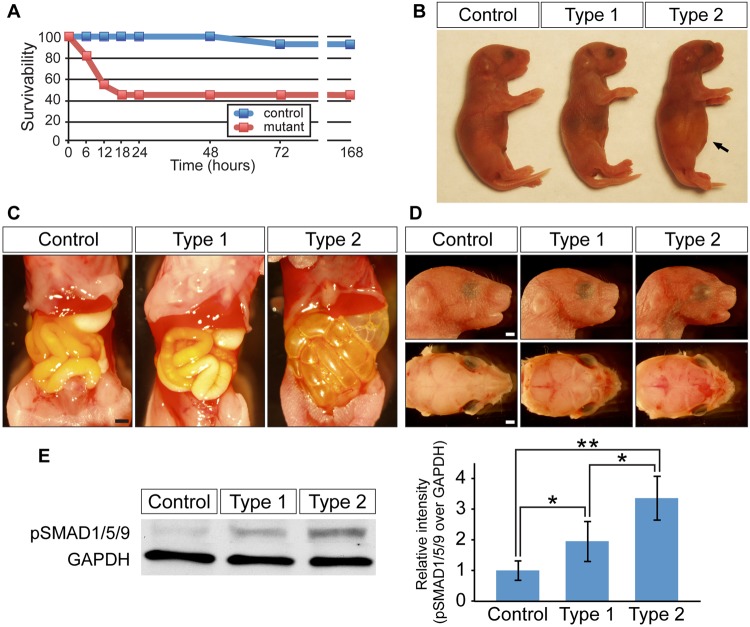
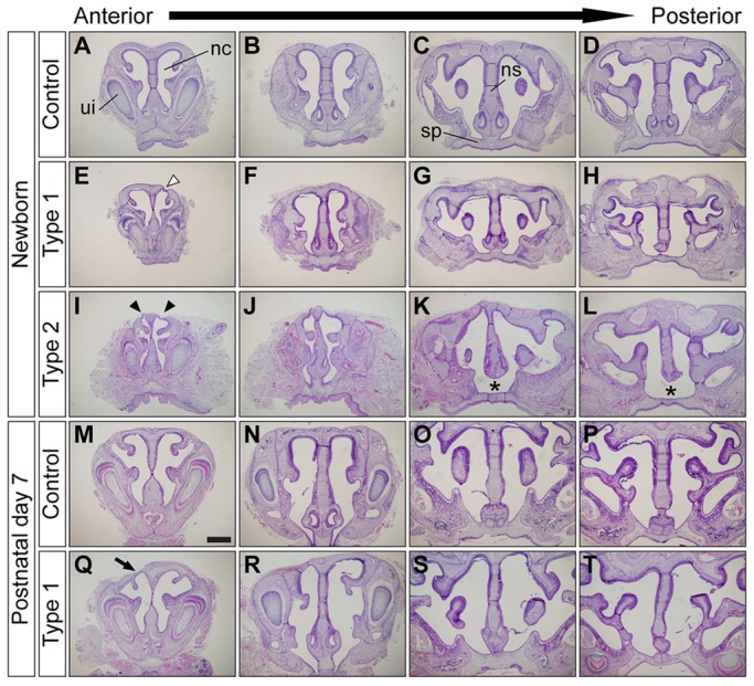
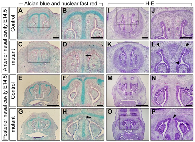
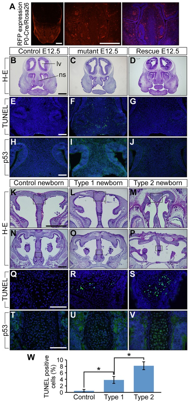
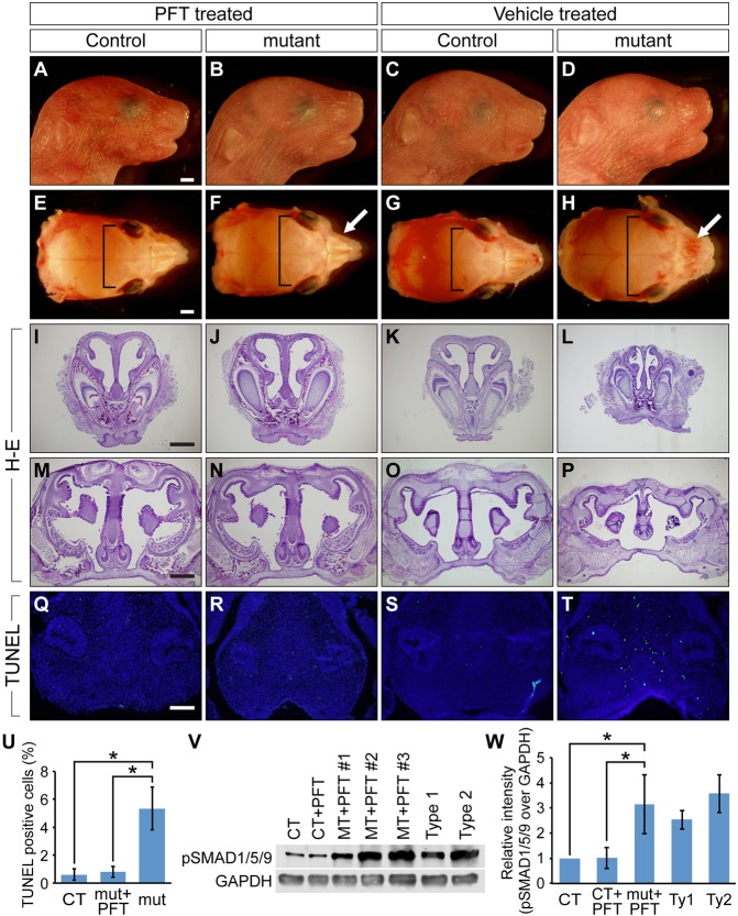
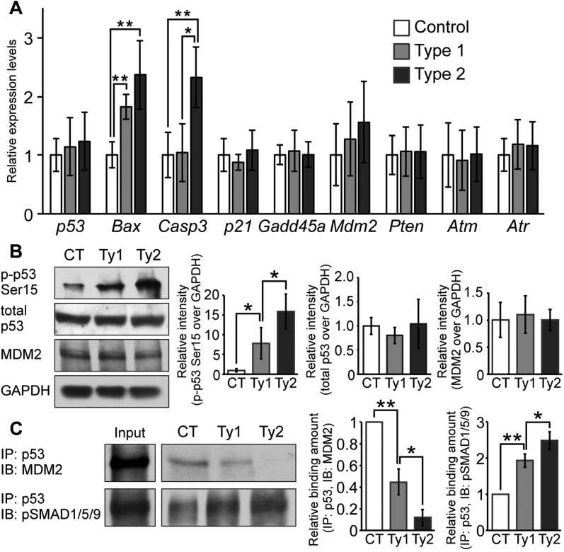
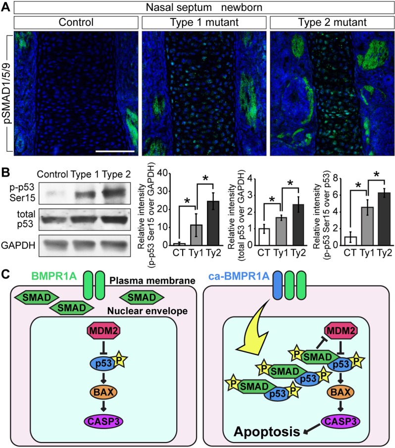
Bone morphogenetic protein (BMP) signaling plays many roles in skull morphogenesis. We have previously reported that enhanced BMP signaling through the BMP type IA receptor (BMPR1A) in cranial neural crest cells causes craniosynostosis during postnatal development. Additionally, we observed that 55% of Bmpr1a mutant mice show neonatal lethality characterized by a distended gastrointestinal tract. Here, we show that severely affected mutants exhibit defective nasal cartilage, failure of fusion between the nasal septum and the secondary palate, and higher levels of phosphorylated SMAD1 and SMAD5 in the nasal tissue. TUNEL demonstrated an increase in apoptosis in both condensing mesenchymal tissues and cartilage of the nasal region in mutants. The levels of p53 (TRP53) tumor suppressor protein were also increased in the same tissue. Injection of pifithrin-α, a chemical inhibitor of p53, into pregnant mice prevented neonatal lethality while concomitantly reducing apoptosis in nasal cartilage primordia, suggesting that enhanced BMP signaling induces p53-mediated apoptosis in the nasal cartilage. The expression of Bax and caspase 3, downstream targets of p53, was increased in the mutants; however, the p53 expression level was unchanged. It has been reported that MDM2 interacts with p53 to promote degradation. We found that the amount of MDM2-p53 complex was decreased in all mutants, and the most severely affected mutants had the largest decrease. Our previous finding that the BMP signaling component SMAD1 prevents MDM2-mediated p53 degradation coupled with our new data indicate that augmented BMP signaling induces p53-mediated apoptosis by prevention of p53 degradation in developing nasal cartilage. Thus, an appropriate level of BMP signaling is required for proper craniofacial morphogenesis.

Keywords: Apoptosis; BMP; Craniofacial development; MDM2; Nasal septum; Neural crest cell; SMAD; p53.


Figures

Fig. 1.
Fig. 1.
Enhanced BMP signaling through a constitutively active form of BMPR1A causes neonatal lethality. (A) Survival curve for control (blue, n=14) and ca-Bmpr1a:P0-Cre mice (red, n=13) after cesarean section at E18.5. (B) Type 2 mutants exhibited abdominal distension (arrow). (C) Gastrointestinal tracts of type 2 mutants were filled with air bubbles, whereas controls and type 1 mutants had milk in their stomachs. (D) Newborn type 1 and type 2 mutants displayed short broad snouts and hypertelorism. (E) Western blot analysis measuring the levels of pSMAD1/5/9 in control, type 1 and type 2 mutant nasal tissues. GAPDH was used as a loading control. Results for pSMAD1/5/9 were quantified by densitometry (bar graph). All data are represented as mean±s.d. (n=4). *P<0.05, **P<0.005 (Student's t-test). Scale bars: 1 mm.
Fig. 2.
Fig. 2.
Augmentation of BMP signaling leads to nasal cartilage defects. (A-L) Frontal sections of newborn control (A-D), type 1 (E-H) and type 2 mutant (I-L) nasal cavities. Type 1 mutants had unilateral nasal cartilage defects (E, white arrowhead). Type 2 mutants had bilateral nasal cartilage defects (I, arrowheads), severe nasal cavity stenosis (I,J) and failure of fusion between the nasal septum and the secondary palate (K,L, asterisks). (M-T) Histological comparison of nasal structures between control (M-P) and type 1 surviving mutants (Q-T) at P7. Frontal sections from four different anteroposterior levels of nasal cavity were stained using Hematoxylin and Eosin (H&E). Type 1 mutants had unilateral nasal cartilage defects (Q, arrow). ui, upper incisor; nc, nasal cavity; ns, nasal septum; sp, secondary palate. Scale bar: 500 µm.
Fig. 3.
Fig. 3.
Inhibition of chondrogenesis in mutants at E14.5. (A-H) Alcian Blue and Nuclear Fast Red staining of E14.5 controls (A,B,E,F) and mutants (C,D,G,H). The level of Alcian Blue-positive mucin was reduced in mutant nasal cartilage (D,H, arrows). (I-P) H&E staining of E14.5 controls (I,J,M,N) and mutants (K,L,O,P). Mutants had a shorter nasal septum (M,O, brackets) and cartilage defects in both anterior and posterior nasal cavity levels (L,P, arrowheads). Boxed areas in A,C,E,G,I,K,M, and O are shown at higher magnification in B,D,F,H,J,L,N and P, respectively. Scale bars: 200 µm in A,B,F,I,J,N; 1 mm in E,M.
Fig. 4.
Fig. 4.
Augmentation of BMP signaling did not change FGF signaling components in the nasal portion of mutant at E14.5. (A) The levels of pERK1/2 were examined using protein lysate from control (CT), type 1 (Ty1), and type 2 mutant (Ty2) nasal tissues. Total ERK1/2 was used as a loading control. (B-U) Immunohistochemistry was performed using antibodies against pERK1/2, FGF2, FGFR1, FGFR2 and FGFR3 (green). The levels of FGF2, FGFR1 and FGFR2 were upregulated in both control and mutant nasal cartilage and mucosal epithelium at E14.5 (F-Q). C,E,G,I,K,M,O,Q,S and U are merged images, with DAPI staining shown in blue. Scale bar: 200 µm.
Fig. 5.
Fig. 5.
Augmentation of BMP signaling leads to increased levels of cell death and p53 protein in developing nasal cartilage of E12.5 mice and newborns. (A) tdTomato distribution in E12.5 P0-Cre transgenic mice crossed with ROSA-tdTomato Cre reporter mice. The right panel is the tdTomato image merged with DAPI staining, which is shown in blue. (B-D) Frontal sections of E12.5 control, mutant and ca-Bmpr1a:P0-Cre:Bmpr1a+/− (rescue) mice were stained using H&E. Control and rescue mice had comparable nasal septum morphology; however, mutants had shorter nasal septa. lv, lateral ventricle; ns, nasal septum. (E-G) TUNEL assay was performed to detect apoptosis. E12.5 mutants exhibited increased numbers of apoptotic cells shown in green (F). Blue indicates DAPI staining. (H-J) Immunohistochemistry using p53 antibody revealed increased p53 levels in mutant nasal septa (I). (K-P) Frontal sections of newborn controls, type 1 and type 2 mutants were stained using H&E. Boxed areas in K-P are shown at high magnification in Q-V, respectively. (Q-S) TUNEL assay was performed to detect apoptosis. Both type 1 and type 2 newborn mutants exhibited more apoptotic cells, which are shown in green (R,S). (T-V) Immunohistochemistry using p53 antibody revealed increased p53 levels in both mutant nasal septa (U,V). (W) Quantification of TUNEL assay results in newborns. All data are represented as mean±s.d. (n=3); *P<0.01 (Student's t-test). Scale bars: 500 µm in A,B,K,N; 100 µm in E,H,Q,T.
Fig. 6.
Fig. 6.
p53-specific chemical inhibition by pifithrin-α rescues nasal tissue defects in vivo. (A-H) Newborn skulls from PFT-treated controls (A,E), PFT-treated mutants (B,F), vehicle-treated controls (C,G) and vehicle-treated mutants (D,H). PFT was intraperitoneally injected to pregnant mice from E8.5 to E18.5. Nasal bone defects found in vehicle-treated mutants were ameliorated by PFT treatment (F,H, white arrows). Hypertelorism found in vehicle-treated mutants was also partially rescued by PFT treatment (F,H, brackets). (I-P) Histological analysis of PFT- and vehicle-treated mice. PFT-treated mutants (J,N) had structurally comparable nasal cartilage to both PFT- and vehicle-treated controls (I,K,M,O), whereas vehicle-treated mutants had nasal cartilage defects (L,P). (Q-T) TUNEL assay in E12.5 PFT- and vehicle-treated mice. PFT was intraperitoneally injected from E8.5 to E11.5. Green indicates TUNEL-positive nuclei and blue indicates DAPI. PFT treatment reduced the number of TUNEL-positive cells. Scale bars: 1 mm in A,E; 500 μm in L,M; 100 μm in Q. (U) Quantification of TUNEL assay. Vehicle-treated mutants showed a significantly higher number of TUNEL-positive cells (n=5). *P<0.005 (Student's t-test). CT, control; mut, mutant. (V) The levels of pSMAD1/5/9 were evaluated by western blotting. Protein lysates were collected from vehicle-treated controls, PFT-treated controls, PFT-treated mutants, vehicle-treated type 1 mutants and vehicle-treated type 2 mutants. GAPDH was used as a loading control. MT, mutant. (W) Results of western blotting were quantified. pSMAD1/5/9 levels of PFT-treated mutants were significantly higher than those of PFT- and vehicle-treated controls (n=3-9 per group). Ty1, type 1 mutant; Ty2, type 2 mutant. *P<0.005 (Student's t-test). All data are represented as mean±s.d.
Fig. 7.
Fig. 7.
Augmentation of BMP signaling leads to p53-mediated apoptosis by preventing MDM2-mediated p53 degradation. (A) Expression levels of p53, Bax, caspase 3, p21, Gadd45a, Mdm2, Pten, Atm and Atr were measured by RT-PCR. Total RNA was isolated from the nasal tissue of newborn controls, type 1 and type 2 mutants (n=4-6 per group). Expression levels of these mRNAs were normalized to Gapdh mRNA. *P<0.01, **P<0.005 (Student's t-test). (B) p-p53-Ser15, total p53 and MDM2 levels were examined by western blotting. GAPDH was used as a loading control. Protein lysate was isolated from nasal tissues of controls (CT), type 1 (Ty1) and type 2 (Ty2) mutants. The levels of p-p53-Ser15 (left), total p53 (center) and MDM2 (right) were normalized to GAPDH and compared between control (CT), type 1 and type 2 mutants (n=4 per group). *P<0.05 (Student's t-test). (C) MDM2-p53 and pSMAD1/5/9-p53 complex formation levels were examined by immunoprecipitation (IP) and immunoblotting (IB). Immunoprecipitation was performed using p53 antibody. MDM2 and pSMAD1/5/9 levels were examined using cell lysates after immunoprecipitation. Input indicates levels of MDM2 or pSMAD1/5/9 in 10% protein lysates before immunoprecipitation. MDM2 (left graph) and pSMAD1/5/9 (right graph) interaction levels were quantified and compared between control, type 1 and type 2 mutants (n=3 per group). *P<0.05, **P<0.005 (Student's t-test). All data are represented as mean±s.d.
Fig. 8.
Fig. 8.
Nuclear accumulation of pSMAD leads to activation of p53 by inhibiting MDM2-mediated p53 degradation in the nasal cartilage. (A) Immunohistochemistry was performed in newborn controls, type 1 and type 2 mutants using pSMAD1/5/9 antibody (green). pSMAD1/5/9 was accumulated in the nuclei of mutant nasal septal chondrocytes. These images were taken by confocal laser-scanning microscopy. Blue indicates DAPI staining. Scale bar: 100 µm. (B) p-p53-Ser15, total p53 and GAPDH levels were quantified by western blotting. Protein lysate was isolated from dissected nasal cartilage of controls (CT), type 1 (Ty1) and type 2 (Ty2) mutants. The levels of p-p53-Ser15 were normalized to GAPDH (left) or total p53 (right). The levels of p53 were normalized to GAPDH (center). These protein levels were compared between controls, type 1 and type 2 mutants. The data are represented as mean ± s.d. (n=3 per group). *P<0.05 (Student's t-test). (C) Cartoon depicting BMP signaling activating p53-mediated apoptosis. Nuclear accumulation of pSMAD facilitates its interaction with p53. This p53 stabilization results in inhibition of MDM2-mediated p53 degradation. This event activates the p53-mediated apoptosis pathway in mutant nasal cartilage.

Similar articles

Cited by

References

    1. Anderson R. M., Lawrence A. R., Stottmann R. W., Bachiller D. and Klingensmith J. (2002). Chordin and noggin promote organizing centers of forebrain development in the mouse. Development 129, 4975-4987. - PubMed
    1. Bachiller D., Klingensmith J., Kemp C., Belo J. A., Anderson R. M., May S. R., McMahon J. A., McMahon A. P., Harland R. M., Rossant J. et al. (2000). The organizer factors Chordin and Noggin are required for mouse forebrain development. Nature 403, 658-661 10.1038/35001072 - DOI - PubMed
    1. Badoux D. M. (1966). Framed structures in the mammalian skull. Acta Morphol. Neerlando Scandinavica 6, 239-250. - PubMed
    1. Bakkers J., Hild M., Kramer C., Furutani-Seiki M. and Hammerschmidt M. (2002). Zebrafish DeltaNp63 is a direct target of Bmp signaling and encodes a transcriptional repressor blocking neural specification in the ventral ectoderm. Dev. Cell 2, 617-627 10.1016/S1534-5807(02)00163-6 - DOI - PubMed
    1. Bingham B., Wang R.-G., Hawke M. and Kwok P. (1991). The embryonic development of the lateral nasal wall from 8 to 24 weeks. Laryngoscope 101, 992-997 10.1288/00005537-199109000-00011 - DOI - PubMed

Publication types

MeSH terms