

Endolymphatic hydrops detected by 3-dimensional fluid-attenuated inversion recovery MRI following intratympanic injection of gadolinium in the asymptomatic contralateral ears of patients with unilateral Ménière's disease
- PMID: 25742875
- PMCID: PMC4360748
- DOI: 10.12659/MSM.892383
Endolymphatic hydrops detected by 3-dimensional fluid-attenuated inversion recovery MRI following intratympanic injection of gadolinium in the asymptomatic contralateral ears of patients with unilateral Ménière's disease
Abstract
Background: The aim of this study was to identify the incidence of endolymphatic hydrops using 3-dimensional fluid-attenuated inversion recovery (3D-FLAIR) magnetic resonance imaging (MRI) in the contralateral ear in patients with unilateral Ménière's disease (MD).
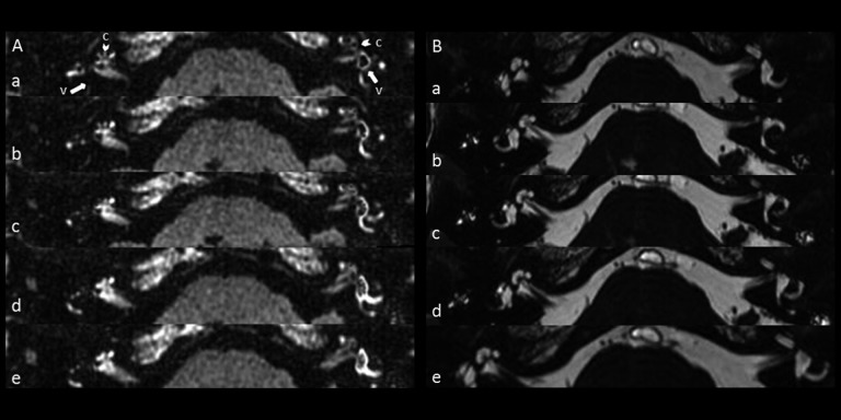
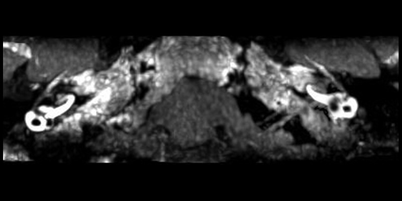
Material and methods: This was a prospective study. 3D-FLAIR MRI was performed with a 3 Tesla (3 T) unit 24 h after the intratympanic administration of gadolinium (Gd) in 30 unilateral MD patients with an asymptomatic contralateral ear. The incidence of contralateral involvement in unilateral MD patients and the potential correlations between the affected and contralateral ears were analyzed.
Results: Endolymphatic hydrops was observed in 7 of the 30 (23.3%) asymptomatic ears. The mean PTA of the asymptomatic ears in the contralateral hydrops patients (33.0±6.1 dB) was significantly higher compared with the non-hydrops patients (17.8±5.7 dB). The patients with observed contralateral hydrops exhibited a significantly longer duration of the disease compared with the non-hydrops patients (6.7±6.3 vs. 2.9±3.1 years, respectively). Furthermore, the patients with contralateral hydrops had a worse hearing level in the affected ears compared with the non-hydrops patients (70.3±7.4 vs. 52.5±3.8 dB, respectively).
Conclusions: Endolymphatic hydrops is closely related to hearing loss but does not necessarily result in Ménière's symptoms. Patients with a long history of MD and severe hearing loss in the affected ear are more likely to exhibit endolymphatic hydrops in the asymptomatic contralateral ear. Adequate attention should focus on unilateral MD patients with contralateral ear hydrops because of the potential to develop bilateral MD.
Figures
Similar articles
-
Reliability of magnetic resonance imaging performed after intratympanic administration of gadolinium in the identification of endolymphatic hydrops in patients with Ménière's disease.Otol Neurotol. 2011 Apr;32(3):472-7. doi: 10.1097/MAO.0b013e31820e7614. Otol Neurotol. 2011. PMID: 21307806
-
[Visualization of endolymphatic hydrops in 3D-FLAIR MRI after intratympanic Gd-DTPA administration in Meniere's disease patients].Zhonghua Er Bi Yan Hou Tou Jing Wai Ke Za Zhi. 2013 Aug;48(8):628-33. Zhonghua Er Bi Yan Hou Tou Jing Wai Ke Za Zhi. 2013. PMID: 24195817 Chinese.
-
Visualization of endolymphatic hydrops and correlation with audio-vestibular functional testing in patients with definite Meniere's disease.Auris Nasus Larynx. 2013 Apr;40(2):167-72. doi: 10.1016/j.anl.2012.07.009. Epub 2012 Aug 4. Auris Nasus Larynx. 2013. PMID: 22867525
-
Novel techniques for the diagnosis of Ménière's disease.Curr Opin Otolaryngol Head Neck Surg. 2013 Oct;21(5):492-6. doi: 10.1097/MOO.0b013e328364869b. Curr Opin Otolaryngol Head Neck Surg. 2013. PMID: 23995329 Review.
-
A perspective from magnetic resonance imaging findings of the inner ear: Relationships among cerebrospinal, ocular and inner ear fluids.Auris Nasus Larynx. 2012 Aug;39(4):345-55. doi: 10.1016/j.anl.2011.05.005. Epub 2011 Aug 25. Auris Nasus Larynx. 2012. PMID: 21871749 Review.
Cited by
-
The Role of Wideband Tympanometry in the Diagnosis of Meniere's Disease.Front Neurol. 2022 Jan 27;13:808921. doi: 10.3389/fneur.2022.808921. eCollection 2022. Front Neurol. 2022. PMID: 35153998 Free PMC article. Review.
-
The clinical features and image characteristics of Meniere's disease patients with endolymphatic hydrops confirmed by enhanced magnetic resonance imaging.Braz J Otorhinolaryngol. 2022 Nov-Dec;88 Suppl 3(Suppl 3):S34-S40. doi: 10.1016/j.bjorl.2021.07.009. Epub 2021 Oct 17. Braz J Otorhinolaryngol. 2022. PMID: 34716108 Free PMC article.
-
Saccular measurements in routine MRI can predict hydrops in Menière's disease.Eur Arch Otorhinolaryngol. 2017 Dec;274(12):4113-4120. doi: 10.1007/s00405-017-4756-8. Epub 2017 Sep 26. Eur Arch Otorhinolaryngol. 2017. PMID: 28951962
-
The Correlation of a 2D Volume-Referencing Endolymphatic-Hydrops Grading System With Extra-Tympanic Electrocochleography in Patients With Definite Ménière's Disease.Front Neurol. 2021 Jan 20;11:595038. doi: 10.3389/fneur.2020.595038. eCollection 2020. Front Neurol. 2021. PMID: 33551957 Free PMC article.
-
Advanced Imaging of the Vestibular Endolymphatic Space in Ménière's Disease.Front Surg. 2021 Aug 23;8:700271. doi: 10.3389/fsurg.2021.700271. eCollection 2021. Front Surg. 2021. PMID: 34497826 Free PMC article. Review.
References
-
- Harris JP, Alexander TH. Current-day prevalence of Meniere’s syndrome. Audiol Neurootol. 2010;15:318–22. - PubMed
-
- Committee on Hearing and Equilibrium guidelines for the diagnosis and evaluation of therapy in Meniere’s disease. American Academy of Otolaryngology-Head and Neck Foundation, Inc. Otolaryngol Head Neck Surg. 1995;113:181–85. - PubMed
-
- Nakashima T, Naganawa S, Sugiura M, et al. Visualization of endolymphatic hydrops in patients with Meniere’s disease. Laryngoscope. 2007;117:415–20. - PubMed
-
- Chen X, Zhang XD, Gu X, et al. Endolymphatic space imaging in idiopathic sudden sensorineural hearing loss with vertigo. Laryngoscope. 2012;122:2265–68. - PubMed
-
- Shimono M, Teranishi M, Yoshida T, et al. Endolymphatic hydrops revealed by magnetic resonance imaging in patients with acute low-tone sensorineural hearing loss. Otol Neurotol. 2013;34:1241–46. - PubMed
Publication types
MeSH terms
Substances
LinkOut - more resources
Full Text Sources
Medical
Miscellaneous
