. 2015 May 1;106(2):237-48.
doi: 10.1093/cvr/cvv097. Epub 2015 Mar 5.

Distinct mPTP activation mechanisms in ischaemia-reperfusion: contributions of Ca2+, ROS, pH, and inorganic polyphosphate

Affiliations

Distinct mPTP activation mechanisms in ischaemia-reperfusion: contributions of Ca2+, ROS, pH, and inorganic polyphosphate

Lea K Seidlmayer et al. Cardiovasc Res. .

Abstract

Aims: The mitochondrial permeability transition pore (mPTP) plays a central role for tissue damage and cell death during ischaemia-reperfusion (I/R). We investigated the contribution of mitochondrial inorganic polyphosphate (polyP), a potent activator of Ca(2+)-induced mPTP opening, towards mPTP activation and cardiac cell death in I/R.

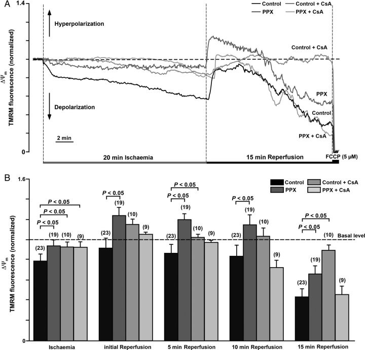
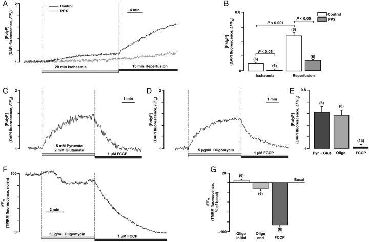
Methods and results: A significant increase in mitochondrial free calcium concentration ([Ca(2+)]m), reactive oxygen species (ROS) generation, mitochondrial membrane potential depolarization (ΔΨm), and mPTP activity, but no cell death, was observed after 20 min of ischaemia. The [Ca(2+)]m increase during ischaemia was partially prevented by the mitochondrial Ca(2+) uniporter (MCU) inhibitor Ru360 and completely abolished by the combination of Ru360 and the ryanodine receptor type 1 blocker dantrolene, suggesting two complimentary Ca(2+) uptake mechanisms. In the absence of Ru360 and dantrolene, mPTP closing by polyP depletion or CSA decreased mitochondrial Ca(2+) uptake, suggesting that during ischaemia Ca(2+) can enter mitochondria through mPTP. During reperfusion, a burst of endogenous polyP production coincided with a decrease in [Ca(2+)]m, a decline in superoxide generation, and an acceleration of hydrogen peroxide (H2O2) production. An increase in H2O2 correlated with restoration of mitochondrial pHm and an increase in cell death. mPTP opening and cell death on reperfusion were prevented by antioxidants Trolox and MnTBAP [Mn (III) tetrakis (4-benzoic acid) porphyrin chloride]. Enzymatic polyP depletion did not affect mPTP opening during reperfusion, but increased ROS generation and cell death, suggesting that polyP plays a protective role in cellular stress response.

Conclusions: Transient Ca(2+)/polyP-mediated mPTP opening during ischaemia may serve to protect cells against cytosolic Ca(2+) overload, whereas ROS/pH-mediated sustained mPTP opening on reperfusion induces cell death.

Keywords: Inorganic polyphosphate; Ischaemia–reperfusion injury; Mitochondrial permeability transition pore; Mitochondrial ryanodine receptor; Oxidative stress.


Figures

Figure 1
Figure 1
(A) Mitochondrial localization of Ca2+-sensitive protein Mitycam (left) was confirmed by colocalization with mitochondrial membrane potential-sensitive dye TMRM (middle) as reflected in the overlay image (right). (B) [Ca2+]m changes measured with Mitycam in permeabilized ventricular myocytes in response to elevation of extramitochondrial [Ca2+] ([Ca2+]em) from 0.1 to 0.8 and 2 µM. Addition of the mitochondrial respiratory chain uncoupler FCCP resulted in release of Ca2+ from mitochondria. (C) Recordings of [Ca2+]m changes monitored with Mitycam, superoxide, and H2O2 generation monitored by MitoSox Red and 2′,7′-dichlorofluorescein, respectively, during I/R in intact control myocytes. (D) Summary of [Ca2+]m (n = 11 cells, four animals), superoxide (n = 11 cells, four animals), and H2O2 (n = 10 cells, three animals) changes during I/R. (E) Recordings of mitochondrial pH (pHm) and redox changes during I/R monitored by genetically encoded mitochondria-targeted probes mito-SypHer and mito-roGFP1, respectively, in intact control myocytes. (F) Summary of mitochondrial pHm (n = 7 cells, three animals) and redox (n = 10 cells, three animals) changes during I/R.
Figure 2
Figure 2
(A and B) Recordings of [Ca2+]m during I/R in control (A, n = 36 cells; 10 animals) and polyP-depleted cells (B, PPX, n = 34 cells; 9 animals) in the presence of Ru360 alone (inhibits MCU; n = 11 cells, three animals in control + Ru360 and n = 7 cells, three animals in the PPX + Ru360 group), Ru360 together with 10 µM dantrolene (inhibits mRyR1, n = 5 cells, three animals in control and n = 7 cells, three animals in the PPX group), CSA (desensitizes mPTP; n = 11 cells, three animals in control + Ru360 and n = 7 cells, three animals in the PPX + Ru360 group), and MnTBAP (superoxide dismutase mimetic, scavenges superoxide; n = 6 cells, three animals in control + MnTBAP and n = 9 cells, three animals in the PPX + MnTBAP group). (C) Average amplitudes for the [Ca2+]m increase at the end of ischaemia and reperfusion in control (black bars) and polyP-depleted (grey bars) cells.
Figure 3
Figure 3
(A and B) Superoxide generation measured with MitoSox Red during I/R in control (A, n = 30 cells, 10 animals) and polyP-depleted (B, PPX, n = 27 cells, 8 animals) cells in the absence and presence of 1 µM Ru360 (n = 10 cells from four animals in control + Ru360; n = 7 cells, three animals in the PPX + Ru360 group), 1 µM CSA (n = 10 cells, three animals in control + CSA; n = 9 cells, three animals in the PPX + CSA group), and 50 µM MnTBAP (n = 7 cells, three animals in control + MnTBAP; n = 8 cells, three animals in the PPX + MnTBAP group). ‘Mock’ ischaemia solution did not contain NaCN (n = 7 cells, three animals in control; n = 8 cells, three animals in the PPX group). (C) Average amplitude of MitoSox Red fluorescence changes at the end of ischaemia and at the end of reperfusion in control and polyP-depleted cells.
Figure 4
Figure 4
Effects of polyP depletion and CSA on mPTP activity and necrotic cell death in I/R. (A) Normalized traces of calcein red fluorescence changes during 20 min ischaemia followed by 15 min of reperfusion in control (n = 23 cells, six animals) and polyP-depleted (PPX, n = 19 cells, eight animals) cells in the absence or presence of 1 µM of CSA (n = 11 cells, three animals for control + CSA; n = 13 cells, four animals for PPX + CSA group). Inserts show images of calcein-loaded cardiomycytes before and after exposure to I/R. (B) Summary of calcein red release from mitochondria (as percentage of basal rate) at the end of ischaemia and reperfusion. (C) Cell death (percentage of LDH release with respect to basal LDH release rates) measured at the end of I/R in control and polyP-depleted (PPX) cells in the absence or presence of 1 µM CSA, 50 µM MnTBAP, 1 µM Trolox (scavenges ROS), and 1 µM Ru360. n is the number of hearts used in each experiment.
Figure 5
Figure 5
(A) Changes in ΔΨm during I/R assessed by TMRM fluorescence in control and polyP-depleted (PPX) cells in the absence or presence of 1 µM CSA. (B) Average values of TMRM fluorescence at the end of ischaemia and at different reperfusion times in control (n = 23 cells, four animals), PPX (n = 19 cells, four animals), control + CSA (n = 10 cells, three animals), and PPX + CSA (n = 9 cells, three animals) groups.
Figure 6
Figure 6
Measurements of polyP levels during I/R. (A) Recordings of DAPI fluorescence in control (with overexpressed GFP) and polyP-depleted (PPX) cardiomyocytes exposed to 20 min of simulated chemical ischaemia followed by a 15 min reperfusion period in normal Tyrode solution. (B) Average values of maximal DAPI fluorescence at the end of 20 min ischaemia and 15 min reperfusion in control (n = 6 cells, three animals) and polyP-depleted (PPX, n = 6 cells, three animals) myocytes. (C) PolyP level changes in control cells upon stimulation of the respiratory chain with 5 mM pyruvate and 2 mM glutamate and subsequent application of 1 µM FCCP. (D) PolyP level changes in control cells upon F1F0-ATP synthase inhibition with 5 µg/mL of oligomycin and subsequent application of 1 µM FCCP. (E) Average values of maximal DAPI fluorescence upon application of oligomycin, pyruvate/glutamate, and FCCP. (F) Changes in ΔΨm during addition of 5 µg/mL of oligomycin and subsequent application of 1 µM FCCP in control cells. (G) Average values of TMRM fluorescence at the beginning and end of oligomycin application and at the end of FCCP application.

Similar articles

Cited by

References

    1. Halestrap AP, Richardson AP. The mitochondrial permeability transition: a current perspective on its identity and role in ischaemia/reperfusion injury. J Mol Cell Cardiol 2015;78:129–141. - PubMed
    1. Halestrap AP. Mitochondria and reperfusion injury of the heart—a holey death but not beyond salvation. J Bioenerg Biomembr 2009;41:113–121. - PubMed
    1. Petronilli V, Penzo D, Scorrano L, Bernardi P, Di Lisa F. The mitochondrial permeability transition, release of cytochrome c and cell death. Correlation with the duration of pore openings in situ. J Biol Chem 2001;276:12030–12034. - PubMed
    1. Perrelli MG, Pagliaro P, Penna C. Ischemia/reperfusion injury and cardioprotective mechanisms: role of mitochondria and reactive oxygen species. World J Cardiol 2011;3:186–200. - PMC - PubMed
    1. Di Lisa F, Ziegler M. Pathophysiological relevance of mitochondria in NAD(+) metabolism. FEBS Lett 2001;492:4–8. - PubMed

Publication types

MeSH terms