. 2015 Feb 22:14:51.
doi: 10.1186/s12943-015-0318-0.

The long noncoding RNA SPRY4-IT1 increases the proliferation of human breast cancer cells by upregulating ZNF703 expression

Affiliations

The long noncoding RNA SPRY4-IT1 increases the proliferation of human breast cancer cells by upregulating ZNF703 expression

Yongguo Shi et al. Mol Cancer. .

Abstract

Background: Long noncoding RNAs (lncRNAs) have emerged recently as a new class of genes that regulate cellular processes, such as cell growth and apoptosis. The SPRY4 intronic transcript 1 (SPRY4-IT1) is a 708-bp lncRNA on chromosome 5 with a potential functional role in tumorigenesis. The clinical significance of SPRY4-IT1 and the effect of SPRY4-IT1 on cancer progression are unclear.

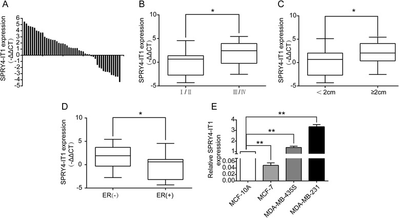
Methods: Quantitative reverse transcriptase PCR (qRT-PCR) was performed to investigate the expression of SPRY4-IT1 in 48 breast cancer tissues and four breast cancer cell lines. Gain and loss of function approaches were used to investigate the biological role of SPRY4-IT1 in vitro. Microarray bioinformatics analysis was performed to identify the putative targets of SPRY4-IT1, which were further verified by rescue experiments, and by western blotting and qRT-PCR.

Results: SPRY4-IT1 expression was significantly upregulated in 48 breast cancer tumor tissues comparedwith normal tissues. Additionally, increased SPRY4-IT1 expression was found to be associated with a larger tumor size and an advanced pathological stage in breast cancer patients. The knockdown of SPRY4-IT1 significantly suppressed proliferation and caused apoptosis of breast cancer cells in vitro. Furthermore, we discovered that ZNF703 was a target of SPRY4-IT1 and was downregulated by SPRY4-IT1 knockdown. Moreover, we provide the first demonstration that ZNF703 plays an oncogenic role in ER (-) breast carcinoma cells.

Conclusions: SPRY4-IT1 is a novel prognostic biomarker and a potential therapeutic candidate for breast cancer.


Figures

Figure 1
Figure 1
Relative expression of SPRY4-IT1 in breast cancer tissues and cells compared with adjacent normal tissues and normal breast epithelial cells. (A) Relative expression of SPRY4-IT1 in breast cancer tissues (T) (n = 48) compared with corresponding non-tumor tissues (N) (n = 48). SPRY4-IT1 expression was examined by qPCR and normalized to GAPDH expression and the levels in the cells of interest were compared with the expression levels inMCF-10A and other cells. The results are presented as the -ΔΔCT changes in tumor tissues relative to normal tissues. (B and C) The data are presented as the relative expression levels in tumor tissues. SPRY4-IT1 expression was significantly higher in patients with a higher pathological stage and a larger tumor size (shown as -ΔΔCT). (D) SPRY4-IT1 expression was assessed by qRT-PCR in ER(−) breast cancer tissues and ER(+) breast cancer tissues(shown as -ΔΔCT). (E) The relative SPRY4-IT1 expression levels in breast cancer cell lines (MD-MB-231, MDA-MB-435S and MCF-7) were compared with human breast epithelial cells (MCF-10A), as assessed by qRT-PCR. * P < 0.05 and **P < 0.01.
Figure 2
Figure 2
Effects of SPRY4-IT1 on breast cancer cell proliferation in vitro. (A) An MTT assay was performed to determine the proliferation of MDA-MB-231, MDA-MB-435S and MCF-7 cells. The data are presented as the means ± S.D. from three independent experiments. (B) Colony-forming growth assays were performed to determine the proliferation of MDA-MB-231, MDA-MB-435S and MCF-7 cells. The colonies were counted and captured. (C) The proliferating MDA-MB-231, MDA-MB-435S and MCF-7 cells were labeled with EdU. The Click-it reaction revealed EdU staining (red). The cell nuclei were stained with Hoechst 33342 (blue). The images are representative of the results obtained. *P < 0.05 and **P < 0.01.
Figure 3
Figure 3
Effect of SPRY4-IT1 on the cell cycle and apoptosis of breast cancer cells in vitro. MDA-MB-231 and MDA-MB-435S cells were transfected with si-SPRY4-IT1 and si-NC, respectively. (A) The bar chart represents the percentage of cells in the G0/G1, S, or G2/M phase, as indicated. The data are presented as the means ± SD from three independent experiments. *P < 0.05. (B) After SPRY4-IT1 knockdown in MDA-MB-231 and MDA-MB-435S cells, the cyclinD1 protein level is diminished compared with the level observed in the control group, as determined by western blot analysis. (C) The percentage of apoptotic cells was determined by flow cytometric analysis. The data represent the means ± SD from three independent experiments. *P < 0.05. (D) Apoptosis in MDA-MB-231 and MDA-MB-435S cells after SPRY4-IT1 knockdown was detected through TUNEL staining. (E) The Bax and Bcl-2 protein levels were elevated in MDA-MB-231 and MDA-MB-435S cells after SPRY4-IT1 expression was blocked compared with the control group.
Figure 4
Figure 4
Gene expression profiling in MDA-MB-231 cells following SPRY4-IT1 knockdown. (A) Clusters of genes altered by SPRY4-IT1 knockdown. The heat map reveals clusters of genes. The green color indicates genes that are up regulated compared with the control cells, and the red color indicates genes that down regulated compared with the control cells. The cells in which SPRY4-IT1 was knocked down are presented as A1 and C1, and the control cells are presented as A2, B2 and C2. (B) Top 10 genes significantly upregulated or down regulated in MDA-MB-231 cells following SPRY4-IT1 knockdown. (C, D) The differential gene expression obtained from the microarray analyses was confirmed by qRT-PCR analysis of 10 selected genes using the gene specific primers shown in Additional file 2: Table S1. The data represent the means of triplicate experiments normalized to the GAPDH level and are presented as the relative fold changes (RFC) of the levels in the MDA-MB-231 cells after SPRY4-IT1 knockdown compared with the levels in the Scr-control cells. (E, F) qPCR analysis of the ZNF703 expression levels in MDA-MB-231 and MDA-MB-435S cells after transfection with scrambled siRNA and si-SPRY4-IT1 (E) and in MCF-7 cells after treatment with an empty vector and pcDNA- SPRY4-IT1 (F). The data are presented as the means ± SD from three independent experiments. **P < 0.01. (G) The ZNF703 protein level is elevated in MDA-MB-231 and MDA-MB-435S cells after transfection with si-SPRY4-IT1 and in MCF-7 cells after transfection with pcDNA-SPRY4-IT1.
Figure 5
Figure 5
Effects of SPRY4-IT1 on ER(−) breast cancer cell proliferation and apoptosis in vitro. (A) The relative ZNF703 expression level in breast cancer cell lines (MDA-MB-231, MDA-MB-435S and MCF-7) compared with the level in human breast epithelial cells (MCF-10A) was assessed by qRT-PCR. (B, C) An MTT assay was performed to determine the proliferation of MDA-MB-231 and MDA-MB-435S cells treated with scrambled siRNA and si-ZNF703. The data are presented as the means ± S.D. from three independent experiments. (D, E) Colony-forming growth assays were performed to determine the proliferation of MDA-MB-231 and MDA-MB-435S cells treated with scrambled siRNA and si-ZNF703. The colonies were counted and captured. (F, G, H) Proliferating MDA-MB-231 and MDA-MB-435S cells treated with scrambled siRNA or si-ZNF703 were labeled with EdU. The Click-it reaction revealed EdU staining (red). The cell nuclei were stained with Hoechst 33342 (blue). The images are representative of the results obtained. (I, J) The percentage of apoptotic cells was determined by flow cytometric analysis. The data are presented as the means ± SD from three independent experiments. (K) An MTT assay was performed to determine the proliferation of MDA-MB-231 cells treated with empty vector and pcDNA-ZNF703. (L, M) Colony-forming growth assays were performed to determine the proliferation of MDA-MB-231 cells treated with empty vector and pcDNA-ZNF703. The colonies were counted and captured. Three independent experiments were performed for each assay. *P < 0.05 and **P < 0.01.
Figure 6
Figure 6
SPRY4-IT1 promotes breast cancer cell proliferation partly through the upregulation of ZNF703 expression. (A) An MTT assay was performed to determine the proliferation of MDA-MB-231 cells transfected with scrambled siRNA, pCDNA-ZNF703,si-SPRY4-IT1 + pCDNA-ZNF703 or si-SPRY4-IT1. (B) An MTT assay was performed to determine the proliferation of MDA-MB-231 cells transfected with scrambled siRNA, si-ZNF703, si-SPRY4-IT1 + si-ZNF703 or si-SPRY4-IT1. The data are presented as the means ± S.D. from three independent experiments. *P < 0.05. (C, D) Colony-forming growth assays were performed to determine the proliferation of MDA-MB-231 treated with scrambled siRNA, pCDNA-ZNF703, si-SPRY4-IT1 + pCDNA-ZNF703, si-ZNF703, si-SPRY4-IT1 + si-ZNF703, or si-SPRY4-IT1. (E, F) Apoptosis in MDA-MB-231 cells treated with scrambled siRNA, si-SPRY4-IT1 + pCDNA-ZNF703, si-SPRY4-IT1 + si-ZNF703, or si-SPRY4-IT1 was detected through TUNEL.

Similar articles

Cited by

References

    1. Gerasimova E, Audit B, Roux SG, Khalil A, Gileva O, Argoul F, et al. Wavelet-based multifractal analysis of dynamic infrared thermograms to assist in early breast cancer diagnosis. Front Physiol. 2014;5:176. doi: 10.3389/fphys.2014.00176. - DOI - PMC - PubMed
    1. Molloy TJ, Devriese LA, Helgason HH, Bosma AJ, Hauptmann M, Voest EE, et al. A multimarker QPCR-based platform for the detection of circulating tumour cells in patients with early-stage breast cancer. Br J Cancer. 2011;104:1913–9. doi: 10.1038/bjc.2011.164. - DOI - PMC - PubMed
    1. Ambrogi F, Biganzoli E, Querzoli P, Ferretti S, Boracchi P, Alberti S, et al. Molecular subtyping of breast cancer from traditional tumor marker profiles using parallel clustering methods. Clin Cancer Res. 2006;12:781–90. doi: 10.1158/1078-0432.CCR-05-0763. - DOI - PubMed
    1. Bosma AJ, Weigelt B, Lambrechts AC, Verhagen OJ, Pruntel R, Hart AA, et al. Detection of circulating breast tumor cells by differential expression of marker genes. Clin Cancer Res. 2002;8:1871–7. - PubMed
    1. Ambrogi F, Fornili M, Boracchi P, Trerotola M, Relli V, Simeone P, et al. Trop-2 is a determinant of breast cancer survival. PLoS One. 2014;9:e96993. doi: 10.1371/journal.pone.0096993. - DOI - PMC - PubMed

Publication types