. 2015 Jun;39(3):306-15.
doi: 10.1007/s00266-014-0439-7. Epub 2015 Mar 6.

Histological characterization of human breast implant capsules

Affiliations

Histological characterization of human breast implant capsules

Janine M Bui et al. Aesthetic Plast Surg. 2015 Jun.

Erratum in

Abstract

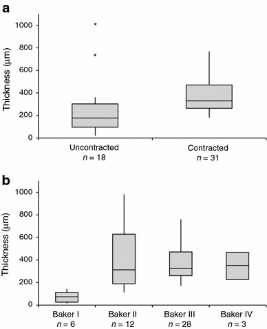
Background: This study investigated the relationships between histomorphological aspects of breast capsules, including capsule thickness, collagen fiber alignment, the presence of α-smooth muscle actin (α-SMA)-positive myofibroblasts, and clinical observations of capsular contracture.

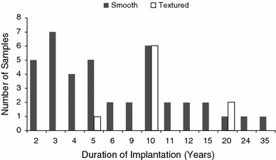
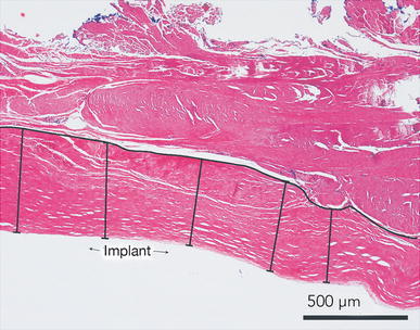
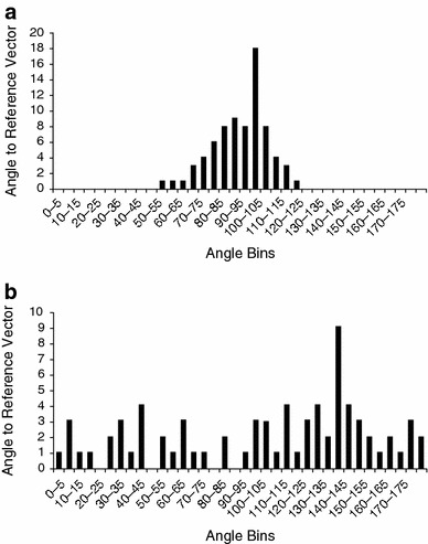
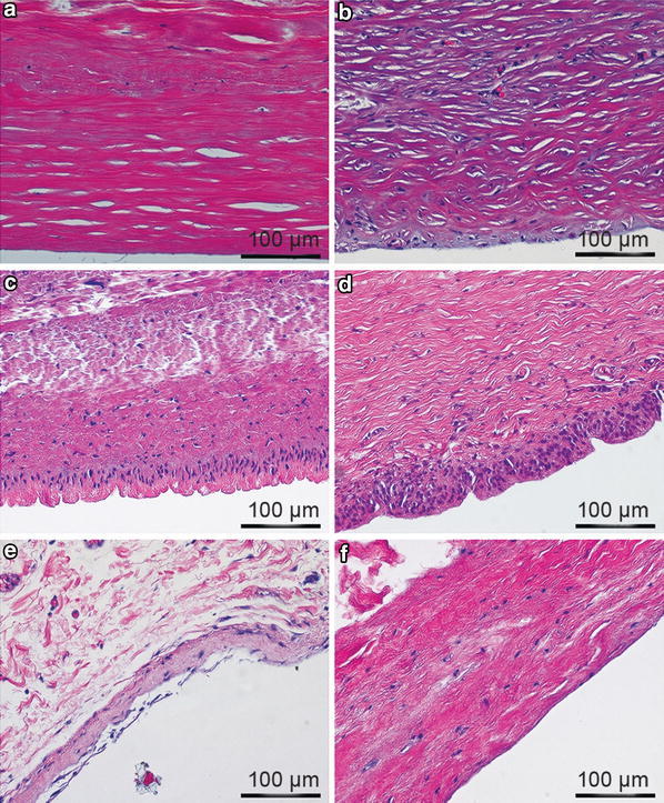
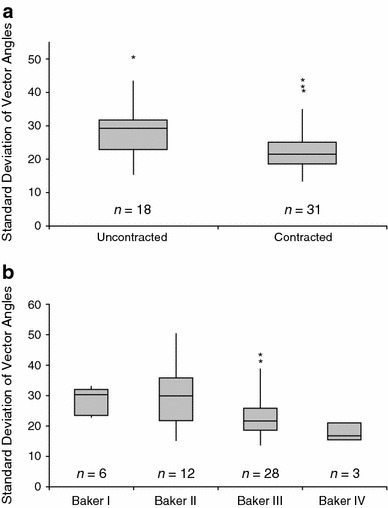
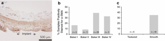
Methods: Breast capsule samples were collected at the time of implant removal in patients undergoing breast implant replacement or revision surgery. Capsular contracture was scored preoperatively using the Baker scale. Histological analysis included hematoxylin and eosin staining, quantitative analysis of capsule thickness, collagen fiber alignment, and immunohistochemical evaluation for α-SMA and CD68.

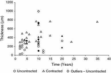
Results: Forty-nine samples were harvested from 41 patients. A large variation in histomorphology was observed between samples, including differences in cellularity, fiber density and organization, and overall structure. Baker I capsules were significantly thinner than Baker II, III, and IV capsules. Capsule thickness positively correlated with implantation time for all capsules and for contracted capsules (Baker III and IV). Contracted capsules had significantly greater collagen fiber alignment and α-SMA-positive immunoreactivity than uncontracted capsules (Baker I and II). Capsules from textured implants had significantly less α-SMA-positive immunoreactivity than capsules from smooth implants.

Conclusion: The histomorphological diversity observed between the breast capsules highlights the challenges of identifying mechanistic trends in capsular contracture. Our findings support the role of increasing capsule thickness and collagen fiber alignment, and the presence of contractile myofibroblasts in the development of contracture. These changes in capsule structure may be directly related to palpation stiffness considered in the Baker score. Approaches to disrupt these processes may aid in decreasing capsular contracture rates.

Level of evidence iii: This journal requires that authors assign a level of evidence to each article. For a full description of these Evidence-Based Medicine ratings, please refer to the Table of Contents or the online Instructions to Authors www.springer.com/00266 .


Figures

Fig. 1
Fig. 1
Summary of patient implants with respect to time from implantation to explant. Duration for smooth implants (n = 40) ranged from 2 to 35 years with an average of 7.9 years, whereas duration for textured implants (n = 9) ranged from 5 to 20 years with an average of 11.7 years. Overall duration averaged 8.6 years for all implants
Fig. 2
Fig. 2
Measurement of capsular thickness. Capsular thickness was measured by drawing a line to delineate the interface between capsule and surrounding tissue where the capsule was defined as the layer of collagenous tissue closest to the implant. Five measurements were taken between the delineating line and the edge of the tissue
Fig. 3
Fig. 3
a Distribution of vectors for a highly aligned capsule with a standard deviation of 13.30 and b a highly unaligned capsule with a standard deviation of 50.21. The distribution of vector angles is representative of fiber alignment and is quantitated by the standard deviation of vectors. If all fibers are parallel, all angles will be either 0° or 180° and the standard deviation of vector angles would be 0. If none of the fibers are parallel, angles will be equally distributed across all measures from 0° to 180°
Fig. 4
Fig. 4
Hematoxylin and eosin staining of human capsules (magnification ×20, scale bar 100 µm). All images are oriented with the implant-tissue interface in the lower portion of the image. a Baker IV contracted capsule with low cellularity and thick dense bands of highly aligned fibers taken from a smooth silicone implant after 3 years of submuscular implantation. b Baker IV contracted capsule with increased cellularity and thick dense bands of highly aligned fibers taken from a smooth silicone implant after 3 years of submuscular implantation. c Baker II capsule with morphology consistent with synovial metaplasia taken from a textured saline implant after 10 years of dual plane implantation. d Baker III capsule with morphology consistent with synovial metaplasia taken from a smooth silicone implant after 15 years of submuscular implantation. e Thin Baker I capsule with loosely arranged fibers taken from a smooth saline implant after 3 years of submuscular implantation. f Baker I capsule with low cellularity and loosely arranged fibers taken from a smooth saline implant after 12 years of subglandular implantation
Fig. 5
Fig. 5
Box plot of capsular thickness by level of contracture. The whiskers represent the minimum and maximum values. The upper and lower edges of the box represent the 25th and 75th percentile, respectively, and the band represents the median. a Contracted capsules (mean = 389.8 µm) are significantly thicker than uncontracted capsules (mean = 285.3 µm; p = 0.0111). Three statistical outliers were identified in the uncontracted group. Outliers included a Baker II capsule from a smooth device that had been implanted for 10 years (thickness = 996 µm), and two Baker II capsules from textured devices that had been implanted for 10 years (thickness = 736 and 723 µm). b Baker I capsules are significantly thinner (mean = 91.5 µm) than Baker II (mean = 408.6 µm; p = 0.0012), III (mean = 393.4 µm; p = 0.0002), and IV capsules (mean = 355.4 µm; p = 0.0282). *Represents statistical outliers
Fig. 6
Fig. 6
Capsular thickness was positively correlated with duration of implantation for all capsules (R 2 = 0.151; p = 0.0076) and for contracted capsules (R 2 = 0.159; p = 0.026), but not for uncontracted capsules (p = 0.296). Solid data points are from textured implants and open data points are from smooth implants. Statistical outliers were only identified in the uncontracted group and were not included in regression analysis. The sample identified at 35 years represents the one patient with breast reconstruction and revision
Fig. 7
Fig. 7
a Box plot of collagen fiber alignment by level of contracture. The whiskers represent the minimum and maximum values. The upper and lower edges of the box represent the 25th and 75th percentile, respectively, and the band represents the median. Contracted capsules (mean = 23.8) had fibers that were significantly more aligned than uncontracted capsules (mean = 29.4; p = 0.0068). b Fiber alignment increased with increasing Baker score (mean Baker scores: I = 30.3, II = 28.9, III = 24.5, and IV = 17.9). One outlier capsule was identified in the Baker II/uncontracted group from a textured device that had been implanted for 10 years (SD = 50.2). Three outliers were identified in the Baker III/contracted group, including a capsule from a textured device that had been implanted for 10 years (SD = 43.3), a capsule from a smooth device that had been implanted for 9 years (SD = 41.1), and a capsule from a smooth device that had been implanted for 2 years (SD = 39.32). *Represents statistical outliers
Fig. 8
Fig. 8
α-Smooth muscle actin (α-SMA) staining of human capsules (magnification ×4, scale bar 500 µm). a Representative α-SMA–positive staining where myofibroblasts can be seen localized to the tissue-device interface. b Percentage of capsules α-SMA–positive for myofibroblasts by Baker score. c Percentage of capsules α-SMA–positive for myofibroblasts by implant surface

References

    1. Adams WP., Jr Capsular contracture: what is it? What causes it? How can it be prevented and managed? Clin Plast Surg. 2009;36:119–126. doi: 10.1016/j.cps.2008.08.007. - DOI - PubMed
    1. Henriksen TF, Fryzek JP, Hölmich LR, et al. Surgical intervention and capsular contracture after breast augmentation: a prospective study of risk factors. Ann Plast Surg. 2005;54:343–351. doi: 10.1097/01.sap.0000151459.07978.fa. - DOI - PubMed
    1. Pajkos A, Deva AK, Vickery K, et al. Detection of subclinical infection in significant breast implant capsules. Plast Reconstr Surg. 2003;111:1605–1611. doi: 10.1097/01.PRS.0000054768.14922.44. - DOI - PubMed
    1. Hwang K, Sim HB, Huan F, Kim DJ. Myofibroblasts and capsular tissue tension in breast capsular contracture. Aesthet Plast Surg. 2010;34:716–721. doi: 10.1007/s00266-010-9532-8. - DOI - PubMed
    1. Abramo AC, De Oliveira VR, Ledo-Silva MC, De Oliveira EL. How texture-inducing contraction vectors affect the fibrous capsule shrinkage around breasts implants? Aesthet Plast Surg. 2010;34:555–560. doi: 10.1007/s00266-010-9495-9. - DOI - PubMed

Publication types