

Hepatitis C virus structural proteins can exacerbate or ameliorate acetaminophen-induced liver injury in mice
- PMID: 25743375
- PMCID: PMC4398656
- DOI: 10.1007/s00204-015-1498-5
Hepatitis C virus structural proteins can exacerbate or ameliorate acetaminophen-induced liver injury in mice
Abstract
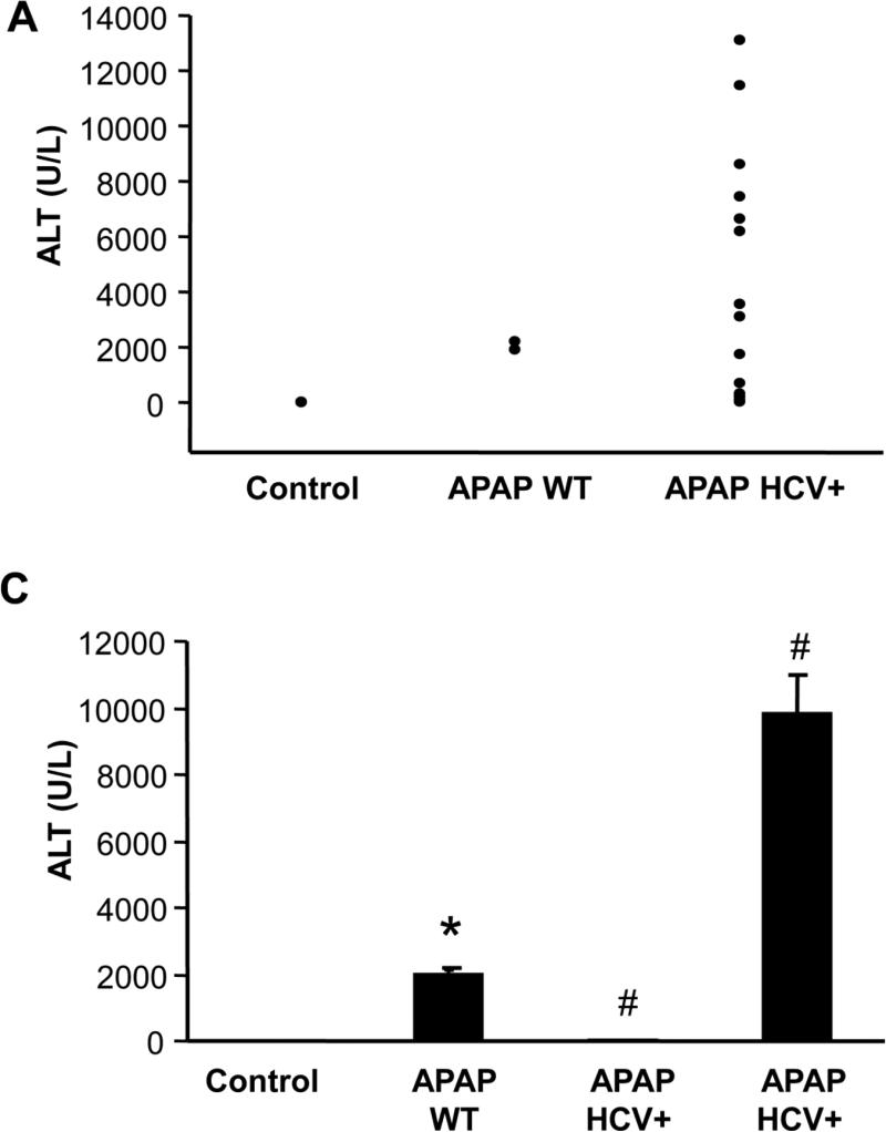
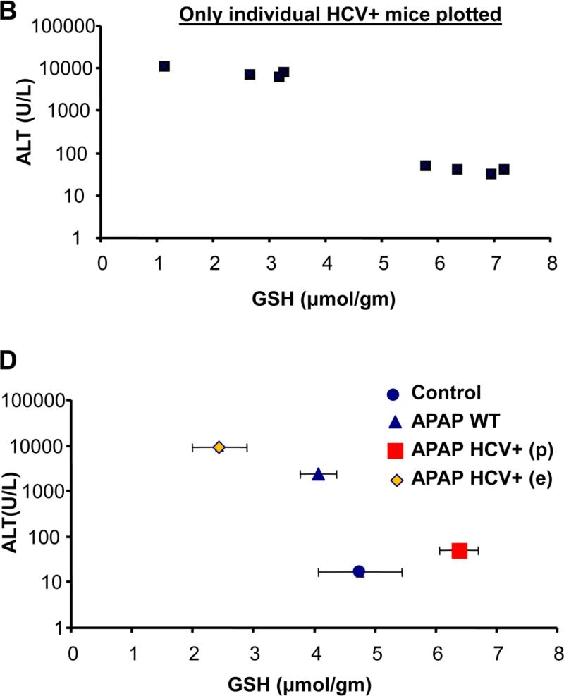
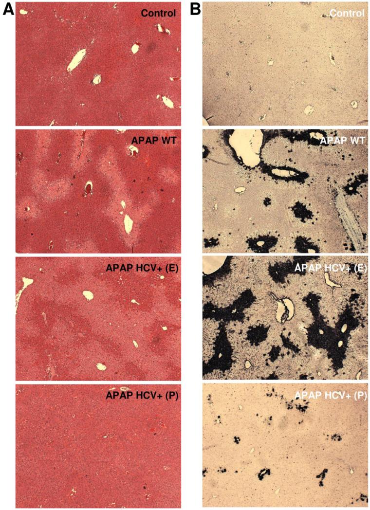
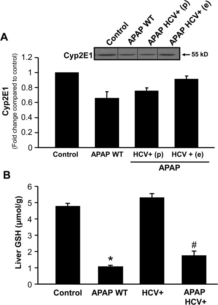
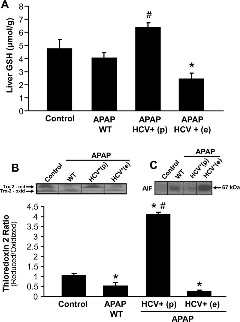
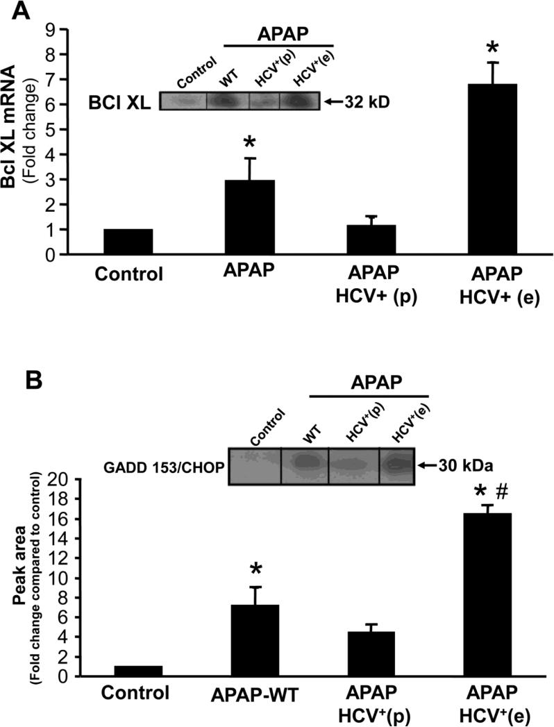
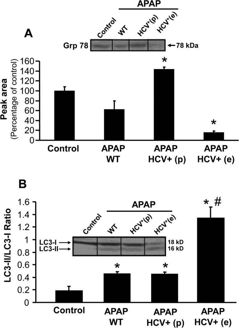
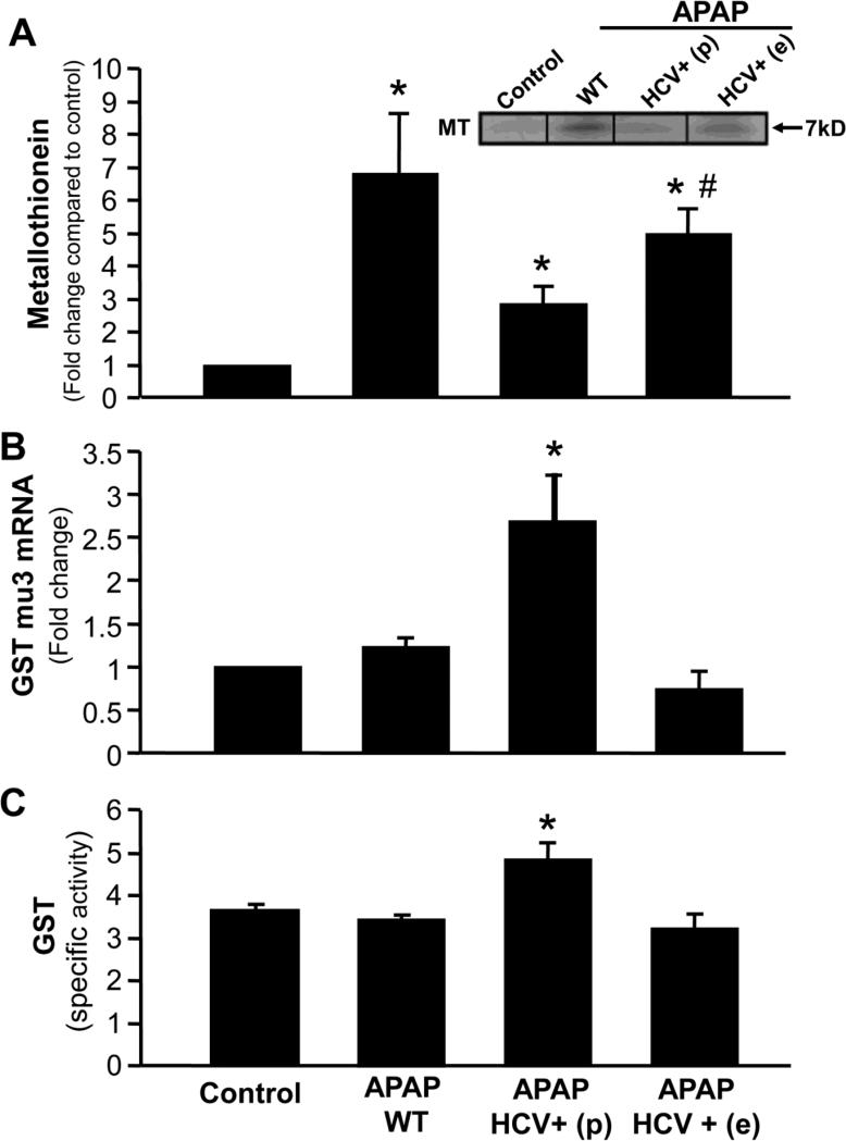
Chronic hepatitis C virus (HCV) infection predisposes patients to develop liver failure after acetaminophen (APAP) overdose. Mechanisms involved in this were explored using transgenic mice expressing the HCV structural proteins core, E1 and E2. Treatment of C57BL/6J mice with 200 mg/kg body weight APAP resulted in significant liver injury at 6 h as indicated by elevated ALT levels, focal centrilobular necrosis and nuclear DNA fragmentation. HCV transgenic mice showed a variable response, with approximately half the animals showing exacerbation of all parameters of liver injury, while the other half was protected. HCV transgenic mice with higher liver injury had lower liver glutathione levels, elevated mitochondrial oxidative stress and enhanced release of apoptosis-inducing factor (AIF) from the mitochondria. This was accompanied by induction of a higher ER stress response and induction of autophagy. Transgenic animals showing protection against liver injury had a robust recovery of liver glutathione content at 6 h when compared to wild-type animals, accompanied by reduction in mitochondrial oxidative stress and AIF release. This was accompanied by an elevation in glutathione S-transferase mRNA levels and activity, which suggests that an efficient clearance of the reactive intermediate may contribute to the protection against APAP hepatotoxicity in these mice. These results demonstrate that while HCV infection could exacerbate APAP-induced liver injury due to induction and amplification of mitochondrial oxidant stress, it could also protect against injury by activation of APAP scavenging mechanisms.
Figures
Similar articles
-
Apoptosis-inducing factor modulates mitochondrial oxidant stress in acetaminophen hepatotoxicity.Toxicol Sci. 2011 Aug;122(2):598-605. doi: 10.1093/toxsci/kfr116. Epub 2011 May 13. Toxicol Sci. 2011. PMID: 21572097 Free PMC article.
-
The impact of partial manganese superoxide dismutase (SOD2)-deficiency on mitochondrial oxidant stress, DNA fragmentation and liver injury during acetaminophen hepatotoxicity.Toxicol Appl Pharmacol. 2011 Mar 15;251(3):226-33. doi: 10.1016/j.taap.2011.01.004. Epub 2011 Jan 15. Toxicol Appl Pharmacol. 2011. PMID: 21241727 Free PMC article.
-
Editor's Highlight: Metformin Protects Against Acetaminophen Hepatotoxicity by Attenuation of Mitochondrial Oxidant Stress and Dysfunction.Toxicol Sci. 2016 Dec;154(2):214-226. doi: 10.1093/toxsci/kfw158. Epub 2016 Aug 25. Toxicol Sci. 2016. PMID: 27562556 Free PMC article.
-
Autophagy and acetaminophen-induced hepatotoxicity.Arch Toxicol. 2018 Jul;92(7):2153-2161. doi: 10.1007/s00204-018-2237-5. Epub 2018 Jun 6. Arch Toxicol. 2018. PMID: 29876591 Review.
-
Acetaminophen Hepatotoxicity: Paradigm for Understanding Mechanisms of Drug-Induced Liver Injury.Annu Rev Pathol. 2024 Jan 24;19:453-478. doi: 10.1146/annurev-pathmechdis-051122-094016. Annu Rev Pathol. 2024. PMID: 38265880 Free PMC article. Review.
Cited by
-
Acetaminophen Hepatotoxicity.Semin Liver Dis. 2019 May;39(2):221-234. doi: 10.1055/s-0039-1679919. Epub 2019 Mar 8. Semin Liver Dis. 2019. PMID: 30849782 Free PMC article.
-
Highlight report: Metabolomics in hepatotoxicity testing.EXCLI J. 2017 Dec 21;16:1323-1325. doi: 10.17179/excli2017-1041. eCollection 2017. EXCLI J. 2017. PMID: 29333135 Free PMC article. No abstract available.
-
Chronic Hepatitis C: Acute Exacerbation and Alanine Aminotransferase Flare.Viruses. 2023 Jan 8;15(1):183. doi: 10.3390/v15010183. Viruses. 2023. PMID: 36680223 Free PMC article. Review.
-
Mixed lineage kinase domain-like protein deficiency exacerbates early injury in a mouse model of acetaminophen hepatotoxicity.Toxicol Sci. 2025 May 1;205(1):220-232. doi: 10.1093/toxsci/kfaf022. Toxicol Sci. 2025. PMID: 39985503
-
HCV core protein represses the apoptosis and improves the autophagy of human hepatocytes.Int J Clin Exp Med. 2015 Sep 15;8(9):15787-93. eCollection 2015. Int J Clin Exp Med. 2015. PMID: 26629077 Free PMC article.
References
-
- Bajt ML, Cover C, Lemasters JJ, Jaeschke H. Nuclear translocation of endonuclease G and apoptosis-inducing factor during acetaminophen-induced liver cell injury. Toxicol Sci. 2006;94:217–225. - PubMed
-
- Bajt ML, Farhood A, Lemasters JJ, Jaeschke H. Mitochondrial bax translocation accelerates DNA fragmentation and cell necrosis in a murine model of acetaminophen hepatotoxicity. J Pharmacol Exp Ther. 2008;324:8–14. - PubMed
-
- Bajt ML, Lawson JA, Vonderfecht SL, Gujral JS, Jaeschke H. Protection against Fas receptor-mediated apoptosis in hepatocytes and nonparenchymal cells by a caspase-8 inhibitor in vivo: evidence for a postmitochondrial processing of caspase-8. Toxicol Sci. 2000;58:109–117. - PubMed
Publication types
MeSH terms
Substances
Grants and funding
LinkOut - more resources
Full Text Sources
Other Literature Sources
Medical
