

Clostridium difficile toxin B intoxicated mouse colonic epithelial CT26 cells stimulate the activation of dendritic cells
- PMID: 25743476
- PMCID: PMC4435672
- DOI: 10.1093/femspd/ftv008
Clostridium difficile toxin B intoxicated mouse colonic epithelial CT26 cells stimulate the activation of dendritic cells
Abstract
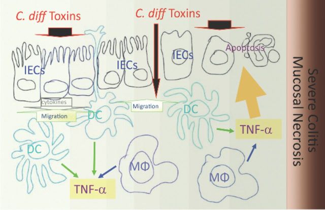
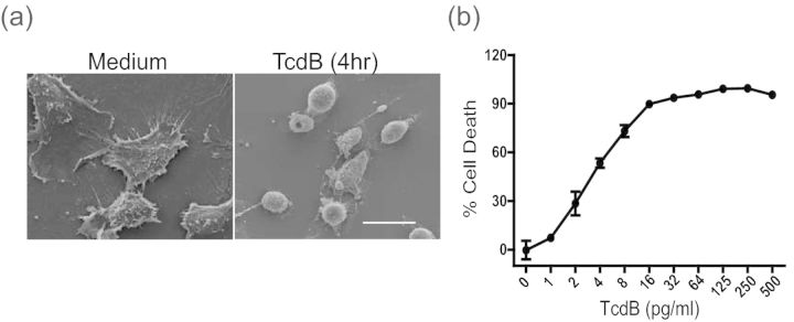
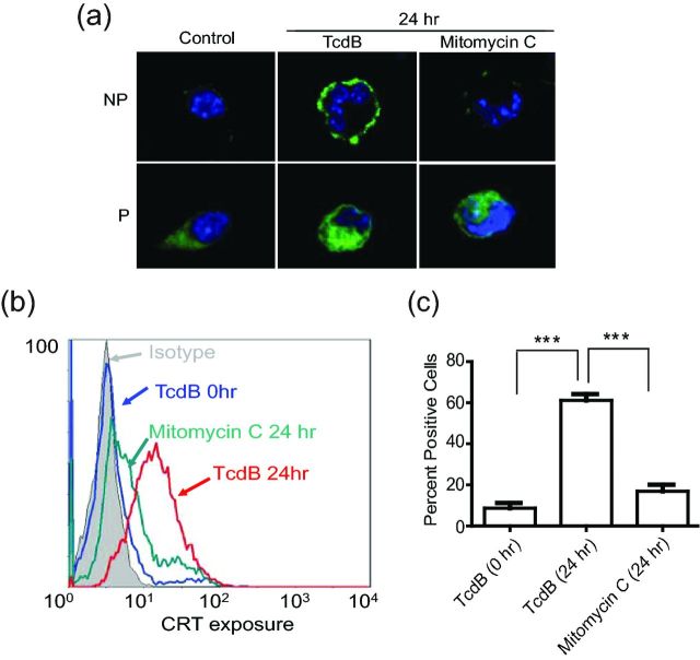
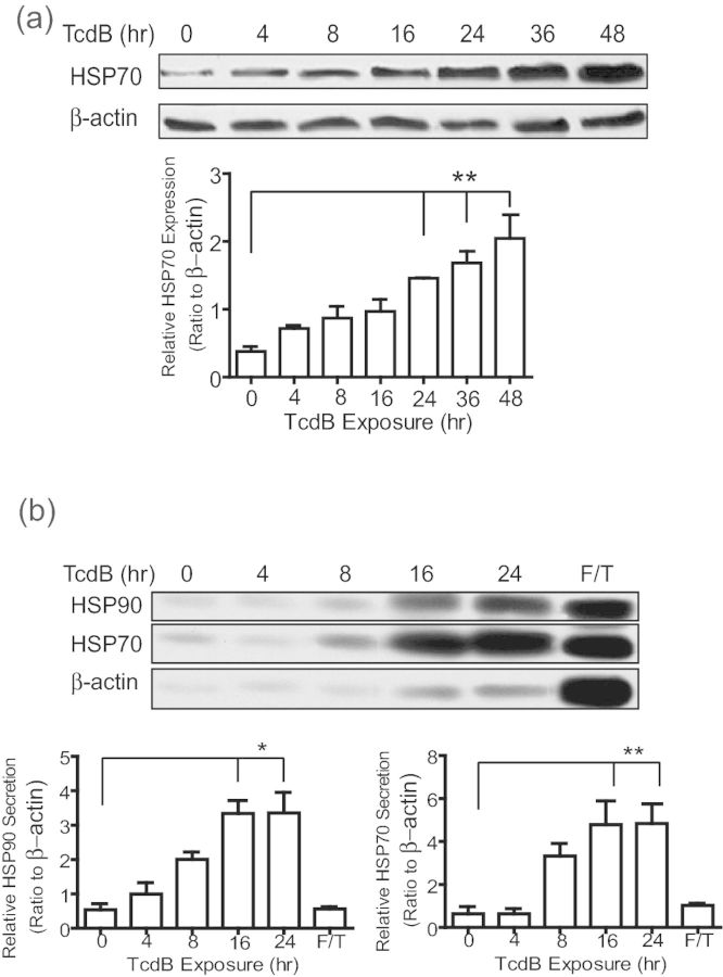
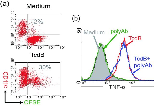
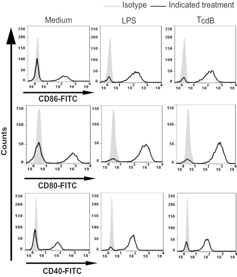
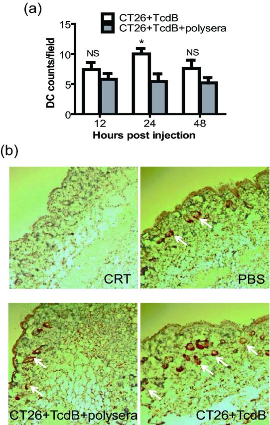
Clostridium difficile causes antibiotic-associated diarrhea and pseudomembranous colitis mainly through two exotoxins TcdA and TcdB that target intestinal epithelial cells. Dendritic cells (DCs) play an important role in regulating intestinal inflammatory responses. In the current study, we explored the interaction of TcdB-intoxicated epithelial cells with mouse bone marrow-derived DCs. TcdB induced cell death and heat shock protein translocation in mouse intestinal epithelial CT26 cells. The intoxicated epithelial cells promoted the phagocytosis and the TNF-α secretion by DCs. Incubation with TcdB-intoxicated CT26 cells stimulated DC maturation. Moreover, TcdB-treated CT26 cells induced DC immigration when they were injected into mice subcutaneously. Taken together, these data demonstrate that TcdB-intoxicated intestinal epithelial cells are able to stimulate DC activation in vitro and attract DCs in vivo, indicating that epithelial cells may be able to regulate DC activation under the exposure of TcdB during C. difficile infection.
Keywords: Clostridium difficile; TcdB; dendritic cells; epithelial cells; inflammation.
© FEMS 2015. All rights reserved. For permissions, please e-mail: journals.permissions@oup.com.
Figures
Similar articles
-
Salubrinal protects against Clostridium difficile toxin B-induced CT26 cell death.Acta Biochim Biophys Sin (Shanghai). 2017 Mar 1;49(3):228-237. doi: 10.1093/abbs/gmw139. Acta Biochim Biophys Sin (Shanghai). 2017. PMID: 28119311
-
Intrarectal instillation of Clostridium difficile toxin A triggers colonic inflammation and tissue damage: development of a novel and efficient mouse model of Clostridium difficile toxin exposure.Infect Immun. 2012 Dec;80(12):4474-84. doi: 10.1128/IAI.00933-12. Epub 2012 Oct 8. Infect Immun. 2012. PMID: 23045481 Free PMC article.
-
High-mobility group box 1 protein contributes to the immunogenicity of rTcdB-treated CT26 cells.Acta Biochim Biophys Sin (Shanghai). 2018 Sep 1;50(9):921-928. doi: 10.1093/abbs/gmy078. Acta Biochim Biophys Sin (Shanghai). 2018. PMID: 30052706
-
Clostridium difficile toxins: mechanism of action and role in disease.Clin Microbiol Rev. 2005 Apr;18(2):247-63. doi: 10.1128/CMR.18.2.247-263.2005. Clin Microbiol Rev. 2005. PMID: 15831824 Free PMC article. Review.
-
Reactive Oxygen Species as Additional Determinants for Cytotoxicity of Clostridium difficile Toxins A and B.Toxins (Basel). 2016 Jan 18;8(1):25. doi: 10.3390/toxins8010025. Toxins (Basel). 2016. PMID: 26797634 Free PMC article. Review.
Cited by
-
The interplay between host immunity and Clostridioides difficile infection.mBio. 2025 Aug 13;16(8):e0356224. doi: 10.1128/mbio.03562-24. Epub 2025 Jul 1. mBio. 2025. PMID: 40590563 Free PMC article. Review.
-
Exploring the Toxin-Mediated Mechanisms in Clostridioides difficile Infection.Microorganisms. 2024 May 16;12(5):1004. doi: 10.3390/microorganisms12051004. Microorganisms. 2024. PMID: 38792835 Free PMC article. Review.
-
Host Immune Responses to Clostridioides difficile: Toxins and Beyond.Front Microbiol. 2021 Dec 21;12:804949. doi: 10.3389/fmicb.2021.804949. eCollection 2021. Front Microbiol. 2021. PMID: 34992590 Free PMC article. Review.
-
Targeting cancer-associated fibroblast-secreted WNT2 restores dendritic cell-mediated antitumour immunity.Gut. 2022 Feb;71(2):333-344. doi: 10.1136/gutjnl-2020-322924. Epub 2021 Mar 10. Gut. 2022. PMID: 33692094 Free PMC article.
References
-
- Baldwin HM, Ito-Ihara T, Isaacs JD, et al. Tumour necrosis factor alpha blockade impairs dendritic cell survival and function in rheumatoid arthritis. Ann Rheum Dis. 2010;69:1200–7. - PubMed
-
- Carneiro-Filho BA, Souza ML, Lima AA, et al. The effect of tumour necrosis factor (TNF) inhibitors in Clostridium difficile toxin-induced paw oedema and neutrophil migration. Pharmacol Toxicol. 2001;88:313–8. - PubMed
-
- Clarke C, Smyth MJ. Calreticulin exposure increases cancer immunogenicity. Nat Biotechnol. 2007;25:192–3. - PubMed
-
- Cloud J, Kelly CP. Update on Clostridium difficile associated disease. Curr Opin Gastroen. 2007;23:4–9. - PubMed
Publication types
MeSH terms
Substances
Grants and funding
LinkOut - more resources
Full Text Sources
Other Literature Sources
