Review
. 2015 Mar;39(2):73-104.
doi: 10.1053/j.semperi.2015.01.002. Epub 2015 Mar 3.

MRI evaluation and safety in the developing brain

Affiliations
Review

MRI evaluation and safety in the developing brain

Shannon Tocchio et al. Semin Perinatol. 2015 Mar.

Abstract

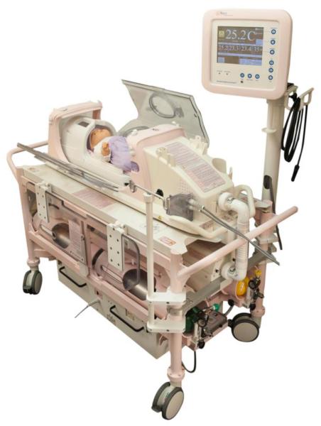
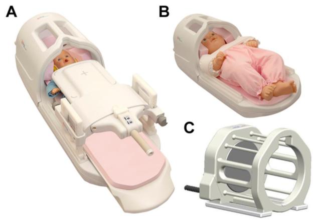
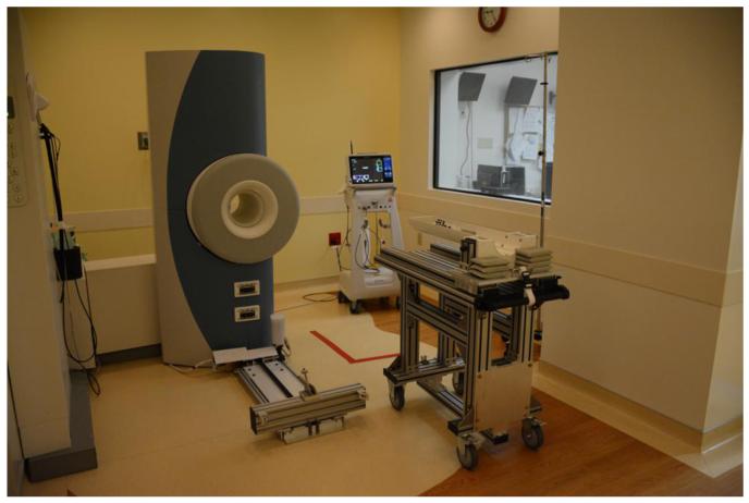
Magnetic resonance imaging (MRI) evaluation of the developing brain has dramatically increased over the last decade. Faster acquisitions and the development of advanced MRI sequences, such as magnetic resonance spectroscopy (MRS), diffusion tensor imaging (DTI), perfusion imaging, functional MR imaging (fMRI), and susceptibility-weighted imaging (SWI), as well as the use of higher magnetic field strengths has made MRI an invaluable tool for detailed evaluation of the developing brain. This article will provide an overview of the use and challenges associated with 1.5-T and 3-T static magnetic fields for evaluation of the developing brain. This review will also summarize the advantages, clinical challenges, and safety concerns specifically related to MRI in the fetus and newborn, including the implications of increased magnetic field strength, logistics related to transporting and monitoring of neonates during scanning, and sedation considerations, and a discussion of current technologies such as MRI conditional neonatal incubators and dedicated small-foot print neonatal intensive care unit (NICU) scanners.

Keywords: MR compatible incubator; MR spectroscopy; NICU magnet; arterial spin label perfusion; fetal MR; neonatal MRI.


Figures

Fig 1
Fig 1. Fetal 3T Imaging and Artifacts
The top row shows two sagittal single-shot fast-spin echo (SSFSE) T2-weighted images obtained on a 3T magnet, demonstrating high resolution anatomic detail of the midline structures in the fetal brain. The bottom row shows two coronal SSFSE images of the fetus demonstrating motion artifact on the left and dielectric artifact on the right, which are both more conspicuous at 3T field strengths and can degrade images of the brain.
Fig 2
Fig 2. Short echo MR spectroscopy in Newborn Brain
T2-weighted sagittal, axial, and coronal MR images of the newborn brain are shown above. The boxes indicate the region of interest (ROI) from where MR spectra will be acquired, with their approximate dimensions provided. The top row of images shows sagittal, axial and coronal images through the left thalamus, with an ROI in the thalamus. At the end of the first row, the spectrum appears normal with no associated abnormality. The bottom row of images shows sagittal, axial and coronal images through the left parietal lobe, with an ROI in the parietal white matter. The spectrum at the end of the row shows an increase in the lactate peak, compatible with hypoxic ischemic injury (HIE). Note that the both ROIs are drawn slightly oblique in order to maximize the sampled volume in the area of interest and avoid partial volume effects. (spectra courtesy of Dr. Stefan Bluml)
Fig 3
Fig 3. ASL perfusion in the newborn brain, patient with recurrent seizures
(A) Gray-scale ASL perfusion image showing two areas of increased perfusion in the region of the peri-Sylvian gyri bilaterally. (B) Corresponding color ASL perfusion image re-demonstrating two foci of increased perfusion in the region of the peri-Sylvian gyri bilaterally. (C) Axial T2-weighted image through the level of the peri-Sylvian gyri corresponding to the region of increased ASL perfusion. (D) Coronal T2-weighted image through the peri-Sylvian gyri corresponding to the region of increased ASL perfusion. Increased perfusion in this case may represent epileptogenic foci in the brain and/or areas of subtle cortical dysplasia.
Fig 4
Fig 4. Neonatal Incubator
The above image shows a second generation SREE MRI conditional, neonate imaging sub-system (NISS-MR). The incubator allows a seamless way to safely bundle, transport and image high-risk infants. Patented, custom built neonatal coils have been integrated into the incubator design allowing high-quality scanning of the infant without removal from the incubator. The NISS-MR consists of an MRI transport incubator, MRI trolley and back-up power supply box (2 hour capacity), which are classified as MR conditional. The incubator is designed to safely accommodate infants up to one month of age, weighing less than 4.5kg or with a whole body length of less than 55cm. The MRI system offers air temperature and humidity regulation as well as the ability to monitor patient skin temperature. The incubator is also designed to accommodate a transport ventilator and wireless pulse-oximeter. (incubator images courtesy of Ravi Srinivasan)
Fig 5
Fig 5. Neonatal Coils
Images of three custom neonatal and infant sized head coils, which can be used to improve signal to noise ratio on examination. (A) Infant Cocoon, which can be used to image infants from 0-6 months of age. (B) Infant Head Spine Array, which can be used to image infants 0-6 months of age. (C) Neonatal Head Coil, which can be used in infants 0-1 months of age. The above coils can all be integrated with the neonatal incubator show in Fig 4. (coil images courtesy of Ravi Srinivasan)
Fig 6
Fig 6. Modified OPTIMA™ MR430s (GE Healthcare, Waukesha, WI) musculoskeletal scanner used at Cincinnati Children’s Hospital Medical Center (CCHMC)
Above is an image of the modified OPTIMA™ MR430s (aka ONI) small-footprint MRI scanner installed at CCHMC. Modifications to the scanner included changing the orientation and height of the magnet, and development of a custom built MRI patient table. The scanner inherently has a smaller, changes to the bore diameter, alteration of the 5-gauss line, decreased magnet weight, decreased cryogen requirements, and improved gradient performance (secondary to smaller gradient RF coils). In addition, control electronics and the radiofrequency system from a state-of-the-art 1.5T GE scanner were integrated with the basic OPTIMA™ MR430s system to give the system the capabilities of a normal department MRI scanner, including advanced imaging techniques, such as MRS, DTI, fMRI, ASL and phase-contrast angiography. The modifications result in feasible placement of a scanner in the NICU and acquisition of high quality diagnostic images.

Similar articles

Cited by

References

    1. US Department of Health and Human Services. Food and Drug Administration. Center for Devices and Radiological Health Criteria for Significant Risk Investigations of Magnetic Resonance Diagnostic Devices - Guidance for Industry and Food and Drug Administration Staff [Internet] 2014 [cited 2014 Sep 3]. Available from: http://www.fda.gov/MedicalDevices/DeviceRegulationandGuidance/GuidanceDo....
    1. Arthur R. Magnetic resonance imaging in preterm infants. Pediatr Radiol. 2006 Jul;36(7):593–607. - PubMed
    1. Van Wezel-Meijler G, Steggerda SJ, Leijser LM. Cranial ultrasonography in neonates: role and limitations. Semin Perinatol. 2010 Feb;34(1):28–38. - PubMed
    1. Miller SP, Ferriero DM, Leonard C, Piecuch R, Glidden DV, Partridge JC, et al. Early brain injury in premature newborns detected with magnetic resonance imaging is associated with adverse early neurodevelopmental outcome. J Pediatr. 2005 Nov;147(5):609–16. - PubMed
    1. Mathur AM, Neil JJ, Inder TE. Understanding brain injury and neurodevelopmental disabilities in the preterm infant: the evolving role of advanced magnetic resonance imaging. Semin Perinatol. 2010 Feb;34(1):57–66. - PMC - PubMed

Publication types