. 2015 Mar 6:6:6302.
doi: 10.1038/ncomms7302.

Genomic landscape of paediatric adrenocortical tumours

Affiliations

Genomic landscape of paediatric adrenocortical tumours

Emilia M Pinto et al. Nat Commun. .

Abstract

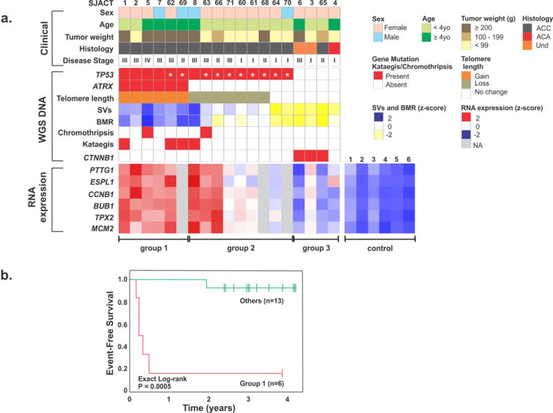
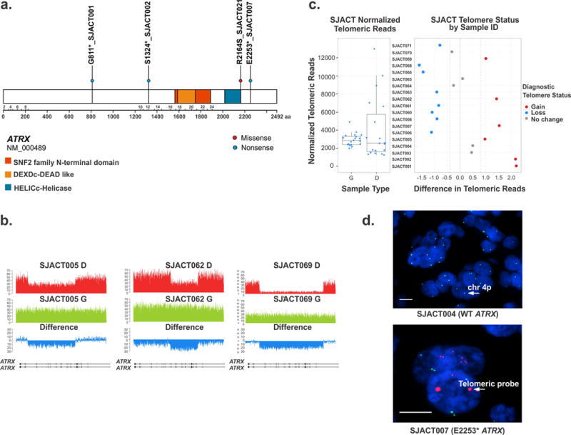
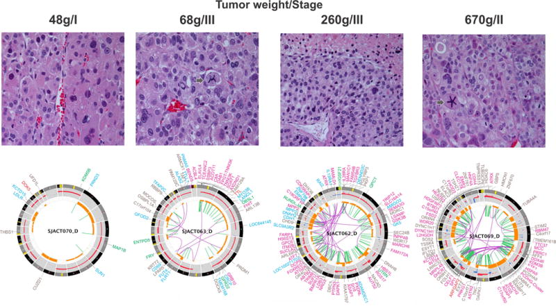
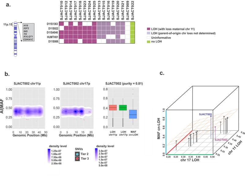
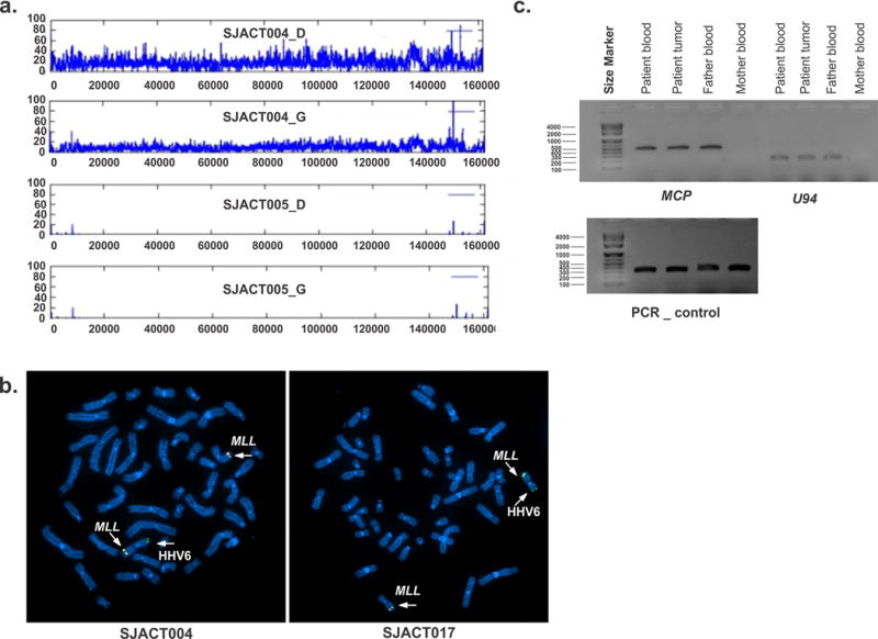
Paediatric adrenocortical carcinoma is a rare malignancy with poor prognosis. Here we analyse 37 adrenocortical tumours (ACTs) by whole-genome, whole-exome and/or transcriptome sequencing. Most cases (91%) show loss of heterozygosity (LOH) of chromosome 11p, with uniform selection against the maternal chromosome. IGF2 on chromosome 11p is overexpressed in 100% of the tumours. TP53 mutations and chromosome 17 LOH with selection against wild-type TP53 are observed in 28 ACTs (76%). Chromosomes 11p and 17 undergo copy-neutral LOH early during tumorigenesis, suggesting tumour-driver events. Additional genetic alterations include recurrent somatic mutations in ATRX and CTNNB1 and integration of human herpesvirus-6 in chromosome 11p. A dismal outcome is predicted by concomitant TP53 and ATRX mutations and associated genomic abnormalities, including massive structural variations and frequent background mutations. Collectively, these findings demonstrate the nature, timing and potential prognostic significance of key genetic alterations in paediatric ACT and outline a hypothetical model of paediatric adrenocortical tumorigenesis.


Conflict of interest statement

Competing financial interests

The authors declare no competing financial interests.

Figures

Figure 1
Figure 1
Association between molecular and clinicopathological features of pediatric adrenocortical tumors. (a) Upper panel: clinicopathological features of 19 patients in the WGS cohort. Center panel: genetic alterations, including mutational status of TP53 (R337H identified by asterisk), ATRX and CTNNB1; telomere length; number of structural variations (SVs); background mutation rate (BMR); chromothripsis and kataegis. Und: undetermined malignancy. Lower panel: RNA expression of selected genes involved in chromosomal segregation and cell cycle control. Three distinct tumor groups (labeled below) emerged from this analysis. Control: normal adrenocortical tissue. (b) Kaplan-Meier probability of event-free survival (exact log-rank test) of pediatric ACT patients in group 1 vs. others.
Figure 2
Figure 2
Somatic ATRX mutations and telomere analysis of pediatric adrenocortical tumors. (a) Distribution of non-silent single nucleotide variations in ATRX (blue dots: nonsense mutations; red dot: missense mutation). (b) Wiggle plots showing internal deletion of multiple exons in ATRX gene in SJACT005, SJACT062, and SJACT069 (diagnostic tumor [D] and germline [G] samples). (c) Relative telomere length determined by WGS in pediatric ACTs vs. matched germline samples. (d) Images of tumor samples hybridized with the telomeric FISH (red) and chromosome 4p (green) probes and stained with DAPI to visualize the nucleus (blue). High magnification views show the large, ultrabright signal in ATRX-mutant adrenocortical tumor cells (SJACT007). Scale bars, 1 μm.
Figure 3
Figure 3
Histopathological and genomic features of TP53-R337H associated ACTs. All tumors had a high mitotic rate (>5 per 50 high-power fields, H&E, 400X). Histology showed a vague nested and trabecular pattern with occasional unpatterned cellular sheets of variable size. Three cases (SJACT063, SJACT062 and SJACT069) had a high nuclear grade and marked cellular pleomorphism. SJACT070 showed occasional enlarged hyperchromatic nuclei with one or more prominent nucleoli. Necrotic cells and atypical mitotic figures (arrow) are identified in SJACT63 and SJACT69. Accumulation of genomic alterations illustrated by the Circos plots (bottom panels) paralleled an increase in tumor weight and correlated with a more aggressive tumor phenotype (Group 1 vs Group 2). Note: labels for gene disrupting SVs (SJACT063 and SJACT062) and non-coding mutations (SJACT069) were removed from the Circos plots.
Figure 4
Figure 4
Characterization and timing of chromosome 11 and 17 LOH in pediatric ACT. (a) Microsatellite analysis of chromosome 11p15 in the WES cohort. All cases with available parental DNA demonstrated selective loss of maternal chromosome 11p15 (n=8, purple). (b) Temporal order of chromosome 11p and 17p cn-LOH and accumulation of single nucleotide variations (SNVs) in SJACT002. Scatter plots show mutant allele fractions (MAFs) of somatic SNVs and their genomic positions (individual dots) combined with 2-D density plots of allelic imbalance (AI) values of germline heterozygous SNPs in cn-LOH regions of chromosomes 11p (left) and 17p (center). At right, AI values in cn-LOH regions of chromosomes 11p and 17p were compared with the MAF distribution of somatic SNVs in genome-wide cn-LOH regions. (c) A 3-D scatter plot summarizes the temporal order of cn-LOH of chromosomes 11p and 17p and somatic SNV accumulation in pediatric ACTs. Shown are median AI values for the chromosome 11p cn-LOH region, median AI values for the chromosome 17p cn-LOH region and median MAF of SNVs in genome-wide cn-LOH regions of 14 cases; SJACT002 and SJACT005 are labeled. See also Supplementary Fig. 7b.
Figure 5
Figure 5
Chromosomal integration of human herpesvirus 6 in pediatric ACT. (a) Coverage plot for cases SJACT004 and SJACT005 showing diagnostic tumor (D) and germline (G) samples reveals integration of the full-length HHV6 genome in SJACT004. The HHV6 genome is duplicated in the tumor sample as a consequence of cn-LOH. (b) Fluorescence in situ hybridization (FISH) of metaphase chromosomes from peripheral blood confirmed HHV6 integration at 11p in both cases. The MLL probe (chromosome 11q23) was used as control (left, SJACT004; right, SJACT017). (c) HHV6 MCP (major capsid protein) and U94 were amplified by PCR from SJACT017 (germline and tumor) and parental DNA demonstrating paternal vertical transmission of integrated HHV6. Exon 6 of the TP53 gene was amplified as DNA quality control.

References

    1. Ribeiro RC, Pinto EM, Zambetti GP, Rodriguez-Galindo C. The International Pediatric Adrenocortical Tumor Registry initiative: contributions to clinical, biological, and treatment advances in pediatric adrenocortical tumors. Mol Cell Endocrinol. 2012;351:37–43. - PubMed
    1. Michalkiewicz E, et al. Clinical and outcome characteristics of children with adrenocortical tumors: a report from the International Pediatric Adrenocortical Tumor Registry. J Clin Oncol. 2004;22:838–845. - PubMed
    1. Wasserman JD, Zambetti GP, Malkin D. Towards an understanding of the role of p53 in adrenocortical carcinogenesis. Mol Cell Endocrinol. 2012;351:101–110. - PMC - PubMed
    1. Weksberg R, Shuman C, Beckwith JB. Beckwith-Wiedemann syndrome. Eur J Hum Genet. 2010;18:8–14. - PMC - PubMed
    1. Steenman M, Westerveld A, Mannens M. Genetics of Beckwith-Wiedemann syndrome-associated tumors: common genetic pathways. Genes Chromosom Cancer. 2000;28:1–13. - PubMed

Publication types

MeSH terms